Table of Contents
What are Cell Organelles?
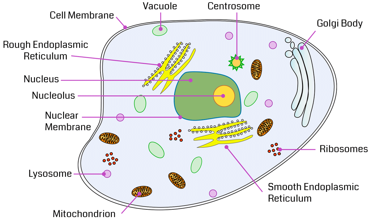
Every single species is composed of a cell which is the basic structural and functional unit of a living organism and is responsible to provide shape, structure and to perform other vital functions. The functional structures suspended in the cytoplasm in a cell are collectively called as cell organelles. They take part in various cellular functions. Cell organelles and components include the various vital components of the cell such as Ribosomes, Endoplasmic Reticulum, Mitochondria, Nucleus, chloroplast, etc.
Cell Organelles – Definition
Cell organelles are the small cellular components present inside the cell. They carry out functions necessary to maintain homeostasis in the cell. It has a particular structural makeup and performs a specific function. These cell organelles have both membrane (layer) and non-membrane(non-film) bound organelles. The cell organelles are involved in many processes like energy production, building proteins and secretions, destroying toxins, and responding to external signals.
Also Check – 10 Important Difference between Cell organelles and Cell Inclusions
Types of cells Organelles
3 Types of Cell Organelles on the basis of cell Membrane
On the basis of presence or absence of membrane, cell organelles are classified into following three types:
Cell Organelles without Membrane
- These cell organelles are not bounded by any membrane.
- They are present in prokaryotic as well as eukaryotic organisms.
- Example: cell wall, Ribosomes, and Cytoskeleton .
Cell Organelles with Single Membranes
- These cell organelles are bounded by a single membrane.
- They are present only in eukaryotic organisms.
- Example:Vacuole, Lysosome, Golgi Apparatus, Endoplasmic Reticulum.
Cell Organelles with Double membranes
- These cell organelles are bounded by a double membrane.
- They are present only in eukaryotic organisms
- Example: Nucleus, Mitochondria and chloroplast .
3 Types of Cell Organelles on the basis of Presence in Plant Cell and Animal Cell
Cell organelles are also divided into three types on the basis of its presence in plant cells and animal cell :
General Cell Organelles
- General cell organelles are the ones which are found in both animal and plant cells.
- Example: cell membrane, reticulum, Golgi apparatus, cytosol, Nucleus, mitochondrion, cytoplasm, lysosome, rough and smooth endoplasmic peroxisome, and the Cytoskeleton.
Temporal Cell Organelles
- Temporal cell organelles are the ones which are found at particular stages of the cell’s life cycle.
- Example: chromosome, the autophagosome, Centrosome, and endosome.
Cell type-specific Cell Organelles
Cell type-specific cell organelles are only found in the plant cells. Example: central Vacuole, chloroplast, and cell wall.
Also Check- Parts of Plant Cell – Location , Structure and Functions
Also Check- 8 Important Differences Between Plant Cell and Animal Cell
List of 24 Important Cell organelles
- Cell membrane / Plasma membrane
- Cell Wall
- Nucleus
- Endoplasmic Reticulum (ER)
- Golgi Apparatus
- Centriole
- Lysosomes
- Microfilaments
- Chloroplast
- Cytoplasm
- Cytoskeleton
- Intermediate filaments
- Plasmodesmata
- Endosomes
- Microtubules
- Storage granules
- Plastids
- Mitochondria
- Vacuole
- Peroxisomes
- Ribosomes
- Cilia and Flagella
- Vesicles
- Microvilli
The Complete guide of Cell Organelles with Definition, Structure, Location, Diagram, Functions and Significance
Cell membrane / Plasma membrane
The plasma membrane is also called a Cell Membrane or Cytoplasmic Membrane.
Cell membrane / Plasma membrane – Definition
The plasma membrane of a cell is defined as a network of lipids and proteins that forms the boundary between a cell’s contents and the outside of the cell.
Structure of Cell membrane / Plasma membrane
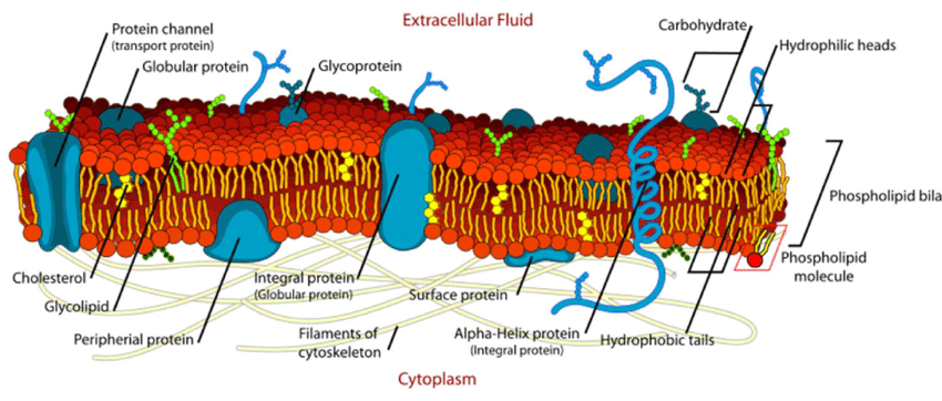
- Plasma Membrane is a type of fluid substance and its structure resembles a fluid mosaic made up of phospholipids, cholesterol, and membrane proteins.
- The fluid mosaic model was coined by S.J. Singer and G.L. Nicolson in 1972.
Components of Cell membrane / Plasma membrane
The components of plasma membrane are discussed below:
Phospholipids
- The membrane is partially made up of molecules called phospholipids which are arranged in a double layer and are called phospholipid bilayer and it is associated with a phosphate group
- It is arranged in such a way that hydrophilic (“water loving”) heads are present on the outside and hydrophobic (“water hating”) tails on the inside. These interactions with water are what allow plasma membranes to form.
Proteins
- Proteins associated with the plasma membrane are either peripheral membrane proteins or integral membrane proteins and act as wedges between the lipids that make up the membrane.
- Peripheral proteins are present either on the internal or external surface of the phospholipid bilayer whereas Integral Proteins are located in the phospholipid bilayer.
- Proteins in the cell membrane play a role in many other functions, such as cell signaling, cell recognition, and enzyme activity.
Carbohydrates
- Plasma membranes also contain carbohydrates; most of these carbohydrates are glycoproteins, the result of carbohydrates attaching to proteins.
- Glycoproteins play a role in the interactions between cells, including cell adhesion, the process by which cells attach to each other.
Cell membrane / Plasma membrane – Diagram
Location of Cell membrane / Plasma membrane
- It is the membrane which surrounds all cells and separates the inner part of the cell from the exterior.
- Plasma membranes also enclose lumens of some cellular organelles.
- It is found in both plant and animal cells.
Functions Cell membrane / Plasma membrane
- It encloses and protects the contents of the cell.
- Provides shape to the cell
- Allows transport of materials in and out of the cell by Diffusion and Osmosis.
- It plays a vital role in both the endocytosis and exocytosis processes.
- It also facilitates communication and signaling between the cells.
- It also maintains the cell potential.
Also Check – Endocytosis -Definition, Mechanism, Types, Examples
SignificanceCell membrane / Plasma membrane
- The plasma membrane, or the cell membrane is a protective cell organelle and is necessary for the protection of a cell.
- It also provides a fixed environment inside the cell. And that membrane has several different functions.
- One is to transport nutrients into the cell and also to transport toxic substances out of the cell.
Also Check – What is plasma Membrane made up of ?
Also Check – Types of Transport Across Cell Membrane
Also Check – 4 Important Components of Plasma Membrane or Cellular Membrane
Also Check 6 Important Functions of Plasma Membrane
Also Check – What would happen if the Plasma Membrane Ruptures or Breakdown?
Also Check – 11 Difference between Cell Wall and Cell Membrane (Plasma Membrane)
Cytoplasm
Cytoplasm Definition-
The cytoplasm is a Colloidal, Viscous, Jelly like fluid which is present between the cell membrane and Nucleus inside the cell. All the cell organelles are embedded on it except the Nucleus. They contain 80-90% water and many organic and inorganic compounds.
Structure of Cytoplasm
It is composed of about 85 % water, 10 to 15 % proteins, 2 to 4 % lipids, nucleic acids, inorganic salts and polysaccharides in smaller amounts. The cytoplasm is also composed of various organelles, which also form the endomembrane system, and the Cytoskeleton.
Layers of Cytoplasm
The Cytoplasm is additionally divided into two layers:
- Ectoplasm
- Endoplasm
Ectoplasm
- Ectoplasm is situated just below or adjacent to the cell membrane forming the outer layer of the protoplasm.
- Ectoplasm is a non-granulated, less dense, thin and shallow layer.
Endoplasm
- Endoplasm is situated deeper within the cell wherever it surrounds the Nucleus and forms the inner layer of the protoplasm.
- Endoplasm consists of Amino acids, Carbohydrates, Lipids, Enzymes, Water, Inorganic Ions etc.
Location of Cytoplasm
- The cytoplasm is enclosed within the cell membrane as is the case with the other cell components/organelles.
- In eukaryotic cells, the cytoplasm is located between the cell membrane/plasma membrane and the nuclear membrane. In prokaryotes, then, the protoplasm occupies the whole cell surroundings (within the plasma membrane) due to the lack of a real Nucleus.
Functions of Cytoplasm
- It Contains enzymes responsible for all the metabolic activity taking place inside the cell.
- It helps in removal of waste material from the cell
- It helps in cell respiration.
- It provides a medium for the organelles to remain suspended.
- It is a means of transport for genetic material.
- The enzymes in the cytoplasm metabolize the macromolecules into small parts so that it can be easily available for the other cellular organelles like Mitochondria.
- It holds organelles in place.
Significance of Cytoplasm
the cytoplasm is made of components that benefit the cell and keep the organelles separate from one another. This helps to keep the cells stable. It is very important for cellular division, cell growth and cell movement.
Also Check – 12 Important Functions of Cell Wall
Also Check – What is Endoplasmic Reticulum ?
Also Check – Parts of Plant Cell – Location , Structure and Functions
Also Check – 8 Important Differences Between Plant Cell and Animal Cell
Nucleus
Nucleus Definition
The Nucleus is the largest, double-membraned organelle found in all eukaryotic cells and acts as the control center of the cellular activities and is the storehouse of the cell’s DNA. It also contains DNA which is the genetic material of an organism.
Structure of Nucleus
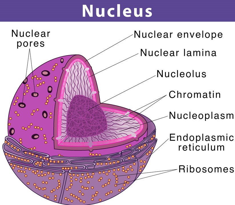
- The Nucleus is a dark, round structure, surrounded by a nuclear membrane. The Nucleus has about 4-6 m diameter. It is present in all eukaryotic cells.
- It is a porous membrane (like cell membrane) and forms a wall between cytoplasm and Nucleus. It Contains DNA, RNA, Protein, nucleolus, and Chromatin network.
- The structure of a Nucleus consists of the nuclear membrane, nucleoplasm, chromosomes, and nucleolus.
Nuclear Membrane
- It is a double-layered structure that encloses the contents of the Nucleus. It differentiates the nuclear constituents from the cytoplasm.
- It also helps in maintaining the shape of the Nucleus of the cell. It consists of nuclear pores which regulates the flow of molecules into and out of the Nucleus .
Nucleoplasm
- Nucleoplasm is also called karyoplasm ,is the gelatinous substance within the nuclear envelope.
- It is composed mainly of water with dissolved salts, enzymes, and organic molecules suspended within.
- It encloses the nucleolus and chromosomes and provides protection to the contents of the Nucleus.
Nucleolus
- It is a dense, membrane-less structure composed of RNA and proteins present within the Nucleus.
- The nucleolus helps to synthesize Ribosomes by transcribing and assembling ribosomal RNA subunits.
Chromosomes
- Chromosomes are present inside the Nucleus and consist of DNA. Chromosomes are present in the form of strings of DNA and histones (protein molecules) called chromatin.
- The Nucleus also contains a number of other non-membrane-delineated bodies like Cajal bodies, Gemini of coiled bodies, polymorphic interphase karyosome association (PIKA), promyelocytic leukemia (PML) bodies etc.
Nucleus – Diagram
Location of Nucleus
In the animal cell, the Nucleus is present in the center of the cell. However, in the plant cell, the Nucleus is present towards the periphery because of the huge water-filled Vacuole which is present in the center.
Functions of Nucleus
- Nucleus Regulates cell functions.
- It controls all metabolic activities of the cell.If removed, the cell dies.
- Nucleus contains chromosomes (bearers of genes that control hereditary characters).It helps in the transmission of hereditary traits from the parent to offspring.
- Nucleus plays an important role in cellular reproduction. In cellular reproduction the cell divides to form two new cells.
- Nucleus Determines the cell development and maturity by directing the chemical activities of the cell.
Significance of Nucleus
- The cell Nucleus is regarded as the most prominent organelle when compared to other cell organelles because it accounts for approximately 10 per cent of the total volume of the cell.
- It is also the storehouse of genetic material DNA and controls the characters and functions of cellsmito in our body.
Also check – 5 Important Functions of Nucleus
Endoplasmic Reticulum
Endoplasmic Reticulum – Definition
The Endoplasmic Reticulum (ER) is a large organelle composed of a network of membranous canals filled with fluid near the Nucleus and stretched throughout the cell. The ER acts as a manufacturing and packaging system.
Structure of Endoplasmic Reticulum
- The structure of the Endoplasmic Reticulum is shaped like a sac.
- The Endoplasmic Reticulum membrane is 50 to 60 A° thick and fluid-mosaic, similar to the plasma membrane unit membrane and is continuous with the plasma membrane, nuclear membrane, and Golgi apparatus membranes.
- The ER membrane system can be anatomically divided into two structures – cisternae and sheets.
- Cisternae are structurally tubular and form a three polygonal-dimensional network. ER sheets are membrane-enclosed, two-dimensional flattened sacs, which extend throughout the cytoplasm.
- The Endoplasmic Reticulum cavity is well developed and serves as a passage for secretory products.
2 Types of Endoplasmic Reticulum (ER)
There are two types of Endoplasmic Reticulum each with distinguishing features:
- Rough Endoplasmic Reticulum
- Smooth Endoplasmic Reticulum
Rough Endoplasmic Reticulum
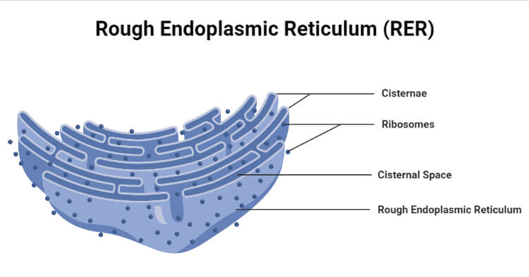
- Rough Endoplasmic Reticulum are considered as “rough” due to the attachment of Ribosomes to the ER membrane.
- Rough Endoplasmic Reticulum are composed of cisternae, tubules, and vesicles.
- Rough Endoplasmic Reticulum synthesizes and secretes proteins in the liver, hormones and other substances in the glands.
- Rough ER is prominent in cells where protein synthesis happens (such as hepatocytes)
Smooth Endoplasmic Reticulum
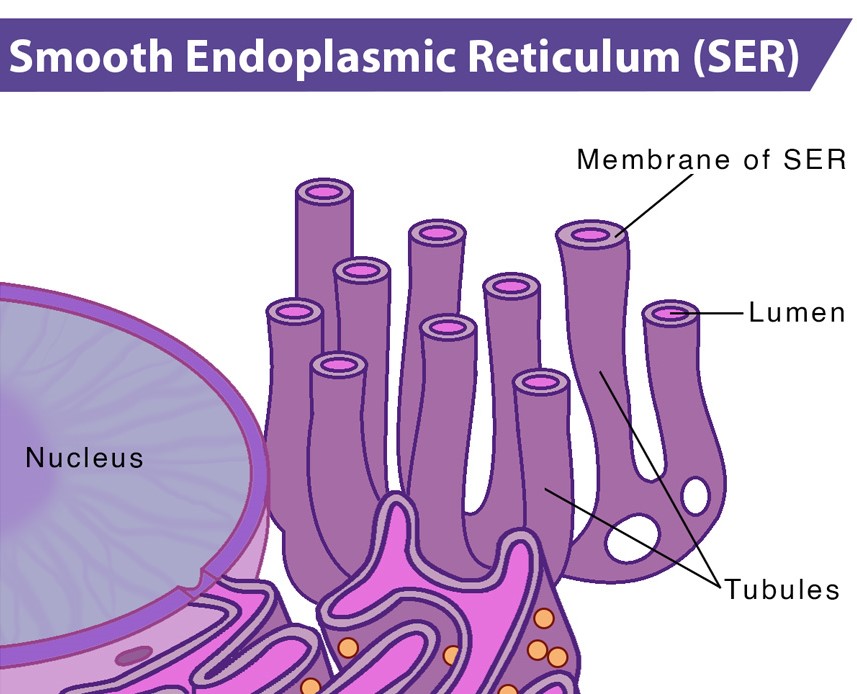
- Smooth Endoplasmic Reticulum are considered as “smooth” due to the absence of Ribosomes on the ER membrane.
- The smooth Endoplasmic Reticulum has a tubular form.
- Smooth Endoplasmic Reticulum acts as a storage organelle for lipid and steroid formation and storage.
- Smooth Endoplasmic Reticulum is also responsible for detoxifying the cell.
Endoplasmic Reticulum- Location
- Endoplasmic Reticulum (ER) is found in all eukaryotic cells.
- In animal cells, the ER usually comprises more than half of the membranous content of the cells.
- The Endoplasmic Reticulum is located near the Nucleus of the cell, where protein synthesis is carried out as a result of DNA translation/transcription.
Endoplasmic Reticulum Functions
- Forms the skeletal framework of the cell.
- It is responsible for the transport of materials from one cell to another.
- Takes part in membrane biogenesis
- It also has a role in detoxification of harmful substances in the liver.
- It serves as an important site for the processing of lipids (fats) and other proteins.
- They aid in nuclear membrane formation during cell division.
- They also provide cellular reactions with increased surface area.
- They play an important role in protein, lipid, glycogen, and other steroid synthesis, such as cholesterol, progesterone, testosterone, etc.
- The ER secretes calcium ions, which are required by the nervous and muscular systems.
Endoplasmic Reticulum Significance
- The Endoplasmic Reticulum (ER) provides around 50 percent of the total membrane surface in an animal cell.
- It plays a major role in the production, processing, and transport of proteins and lipids.
Also Check – 6 Important Difference between Rough and Smooth Endoplasmic Reticulum
Also Check – 7 Difference Between Mitochondria and Endoplasmic Reticulum
Also Check – 11 Important Functions of Endoplasmic Reticulum (ER) Class 9
Also Check – 16 Important Differences between Prokaryotic and Eukaryotic cell
Mitochondria
Mitochondria- Definition
Mitochondria are the oxygen-consuming ribbon-shaped double-membraned organelles floating free throughout the cell. Mitochondria is also called the powerhouse of the cell as they supply all the necessary biological energy to the cell.
Structure of Mitochondria
Mitochondria are mobile, plastic organelles with a diameter of 0.5 to 1.0 micrometer.
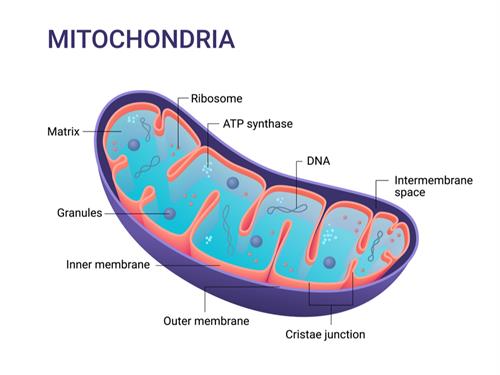
4 Distinct Parts of Mitochondria
- The outer membrane
- The inner membrane
- The intermembrane space
- The matrix.
The Outer Membrane
- It is very similar to the plasma membrane in terms of structure and chemical composition.
- The outer membrane has pores which allow small molecules to be exchanged between the cytoplasm and the intermembrane space.
The Inner Membrane
- This membrane is rich in many enzymes, coenzymes, and other components of the electron transport chain.
- It has finger-like outgrowths called cristae towards the lumen of the mitochondrion and contains tennis-racket shaped F1 particles that contain ATP-ase enzyme for ATP synthesis.
Intermembrane space
- The space between the outer and inner membrane makes up the intermembrane space.
- It has the same composition as that of the cell’s cytoplasm.
The Mitochondrial matrix
It is the gel-like (colloidal) area enclosed by the inner membrane and is composed of soluble enzymes of the Krebs cycle which completely oxidize the acetyl-CoA to produce CO2, H2O and hydrogen ions.
The matrix also contains single or double circular and double-stranded DNA molecules called mtDNA and also the 55S Ribosomes, called mitoRibosomes.
Mitochondria – Diagram
Mitochondria -Location
They are abundantly found on those sites where energy is earnestly required such as sperm tail, muscle cell, liver cell (up to 1600 Mitochondria), microvilli, oocyte (more than 300,000 Mitochondria), etc.
Functions of Mitochondria
- The most crucial function of Mitochondria is to produce energy to drive a host of cellular reactions and mechanisms.
- It takes part in the process of oxidative phosphorylation.
- It also helps the cells to maintain the proper concentration of calcium ions within the compartments of the cell.
- Helps in detoxifying ammonia in the liver cells
- Responsible for building certain parts of the blood and various hormones like testosterone and estrogen
Significance of Mitochondria
- There are about 2000 Mitochondria per cell, representing around 25% of the cell volume.
- They are the PowerHouse of The Cell / Storage Batteries.
- It provides energy by breaking down carbohydrate and sugar molecules to drive a host of cellular reactions and mechanisms.
Also Check – Why is the Mitochondria called the Powerhouse of the Cell
Also Check – 10 Important Functions of Mitochondria
Also Check – 9 Important Difference Between Golgi Bodies and Mitochondria
Also Check – 16 Important Differences between Prokaryotic and Eukaryotic cell
Plastids
Definition- Plastid is a double membrane-bound organelle containing pigments involved in the synthesis and storage of food found within the cells of photosynthetic plants. They are found in plants and algae.
Structure of Plastid
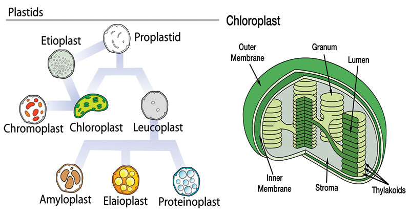
There are different types of Plastids with different structural differences:
- Chloroplasts
- Chromoplasts
- Leucoplasts
- Gerontoplasts
Chloroplasts
- Chloroplasts are biconvex shaped, semi-porous, green coloured, double membranes, cell organelle found within the mesophyll of the plant cell.
- They are usually 4-6 µm in diameter .
- The Chloroplast is bounded by two lipoprotein membranes, an outer and an inner membrane, with an intermembrane space between them.
- Chloroplasts are responsible for photosynthesis.
- The chloroplast is filled with thylakoids, which is where photosynthesis occurs, and chlorophyll remains.
Chromoplasts
- They are blue, red, yellow Coloured Plastid. The Chloroplasts actually convert over to Chromoplasts.
- They contain carotenoid pigments that allow for the different colors seen in fruits and the fall leaves.
Leucoplasts
- Leucoplasts are the non-pigmented organelles found in most of the non-photosynthetic parts of the plant like roots.
- Leucoplasts act as storage sheds for starches, lipids, and proteins depending on the needs of the plants.
- A Leucoplasts may be an amyloplast that stores starch, an amyloplast that stores fat, or a proteinoplast that stores proteins.
Gerontoplasts
- Gerontoplasts are basically Chloroplasts that are going through the aging process.
- It controls the dismantling of the photosynthetic during the senescence of the plant.
Location of Plastids
Plastids are major organelles found in the cells of plants and algae and the cells of various eukaryotic organisms. Plastids are found inside a cell.
Plastids– Diagram
Functions of Plastids
- Plastids play an important role in manufacturing food and storing it.
- Chloroplasts are the centers of synthesis and metabolism of carbohydrates.
- Like Mitochondria, Plastids have their own DNA and Ribosomes. Hence, they may be used in phylogenetic studies.
- Plastids help in the release of oxygen.
- Chromoplast impart color to flowers which help in pollination
- Amyloplast is responsible for the storage of starch.
- Aleuroplast is responsible for the storage of protein.
- Elaioplast is responsible for the storage of fat.
Significance of Plastids
- Plastids usually contain pigments inside them that are used in photosynthesis.
- The variety of a Plastid’s pigments determines the color of the cell.
- Plastid is an important cell organelle that generates energy in plants only.
Also Check – Parts of Plant Cell – Location , Structure and Functions
Also Check – 10 Difference between Mitochondria and Plastids
Ribosomes
Ribosomes – Definition
Ribosomes are tiny spheroidal dense macro-molecular particles composed of RNA and protein that converts genetic code into chains of amino acids. Ribosomes are primarily found in most prokaryotic and eukaryotic cells.
Structure of Ribosome
Ribosome is also known as a ribonucleoprotein as it is a complex of RNA and protein. Ribosomes are made of proteins and ribonucleic acid (abbreviated as RNA), in almost equal amounts.
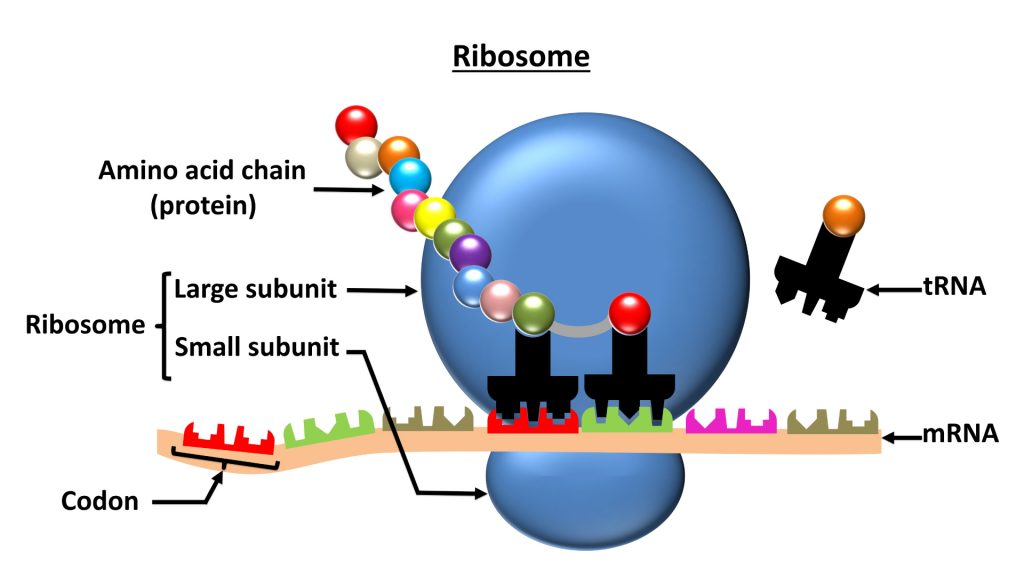
2 Subunits of Ribosomes
Ribosomes are composed of two subunits – smaller and larger, 60S and 40S in eukaryotes both made up of RNA.
- A smaller subunit which binds to a larger subunit and the mRNA pattern.
- A larger subunit which binds to the tRNA, the amino acids, and the smaller subunit.
The Ribonucleic acid is obtained from the nucleolus, at the point where Ribosomes are arranged in a cell.
Ribosomes – Diagram
Location of Ribosomes
- Ribosomes are organelles located inside the animal, human cell, and plant cells.
- They are Situated in two areas of the cytoplasm.
- Usually Scattered in the cytoplasm and a few are connected to the Endoplasmic Reticulum to form rough Endoplasmic Reticulum (ER).
Functions of Ribosomes
- Ribosomes main function is Synthesis of Proteins
- Ribosomes bring together amino acids to form particular proteins, important for completing the cell’s activities.
- By DNA transcription, deoxyribonucleic acid creates mRNA, which is then used to generate proteins.
- During DNA translation, hereditary information from mRNA is translated into proteins.
- Ribosomes are responsible for producing biological proteins.
Significance of Ribosomes
Ribosomes are responsible for protein synthesis which is necessary for the cell’s tasks to be completed. Hereditary information from the mRNA is converted into proteins amid DNA translation.
Golgi Apparatus
Golgi Apparatus, also known as the Shipping Department of Cell. it was discovered by Camillo Golgi in 1898.
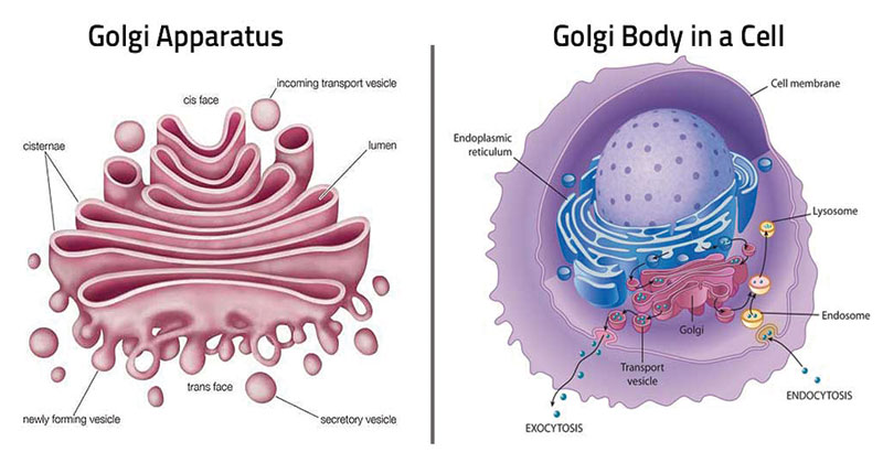
Golgi Apparatus – Definition
Golgi Apparatus or Golgi Complex is a membrane-bound organelle found in eukaryotic cells and is primarily responsible for transporting, modifying, and packaging proteins and lipids to targeted destinations.
Structure of Golgi Apparatus
- The Golgi body contains 5 to 8 cup-shaped, sac-like compartments called cisternae with a diameter of 0.5 µm to 1.0 µm.
- A Golgi stack mostly contains 4 to 8 cisternae.
- The membrane stack of the cisterna divides the interiors from the cytoplasm.
- The Golgi cisternae are placed circularly close to the Nucleus, with discrete convex cis face, also known as forming face, and concave trans, which means maturing face, that is different but yet related, cis face or receiving face and trans face or supplying face.
Golgi Apparatus– Diagram
Location of Golgi Apparatus
Golgi Apparatus is found within the cytoplasm mostly near the Nucleus of a cell and is present in both plant and animal cells.
Functions of Golgi Apparatus
- Golgi Apparatus has an important role in modification, packaging, and transport of materials.
- Golgi Apparatus takes part in synthesis of lysosomes, plasma membranes.
- Golgi Apparatus receives proteins from Endoplasmic Reticulum.
- Golgi Boddies take part in the transport of lipids.
- Golgi Apparatus is the site for the synthesis of various glycolipids, sphingomyelin, etc.
- Sugars are converted into cell wall components by enzymes in the Golgi body in plants.
Significance of Golgi Apparatus
The Golgi also has important functions in tagging vesicles with proteins and sugar molecules, which serve as identifiers for the vesicles so they can be delivered to the proper target.
Also Check – 7 Important Functions of Golgi Apparatus
Also Check – What would happen to the life of a cell if there were no Golgi apparatus ?
Also Check – 9 Important Difference Between Golgi Bodies and Mitochondria
Vacuoles
Vacuoles- Definition
A Vacuole is a membrane-bound cell organelle present in the cytoplasm and filled with a watery fluid containing various substances.
Structure of Vacuoles
- A Vacuole is a membrane bound structure which is composed of phospholipids.
- The membrane surrounding the Vacuole is known as tonoplast.
- The components of the Vacuole are called cell sap.
- The membranes of Vacuoles are embedded with proteins that help in transporting molecules across the membrane.
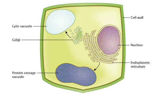
Location of Vacuoles
- Vacuole is a membrane bound structure found in the cytoplasmic matrix of a cell.
- It is found in both animals and plants.
- In animal cells, Vacuoles are generally small and help sequester waste products.
- In plant cells, Vacuoles help maintain water balance.
Vacuoles – Diagram
Functions of Vacuoles
- It provides a shield to the remaining cell part from dangerous molecules and substances.
- The primary function is storage. It stores salts, minerals, pigments and proteins within the cell.
- Vacuoles provide shape to the cell and help it to withstand extreme conditions.
- Vacuoles ensure continued hydrostatic pressure in the cell.
- Vacuoles help to get rid of all the unwanted things inside a cell.
- Vacuoles hold proper pressure in a plant cell, and help in its growth.
Significance of Vacuoles
Vacuoles are the vacant structure serving as the storage organ of the cell. Vacuoles store nutrients and water on which a cell can rely for its survival. They also store the waste from the cell and prevent the cell from contamination.
Also Check – Can a cell live without vacuoles…
Also Check – What is the Difference between Plant Vacuoles and Animal Vacuoles?
Also Check – 10 Important Vacuoles Functions
Also Check – Why do Plant cells possess Large Sized Vacuoles ?
Microbodies
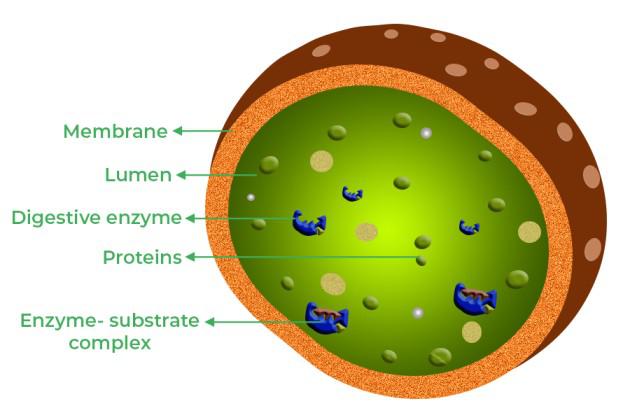
Microbodies – Definition
Microbodies are single membrane bound globular shaped organelle found in eukaryotic cells. They are degradative enzymes that are encapsulated within a single membrane and are considered as containers for metabolic activity.
Structure of Microbodies
- Microbodies are called cytosomes due to their presence in the cytosol of the cell.
- A Microbody is usually a vesicle with a spherical shape and its diameter ranges from 0.2-1.5 micrometers.
- Microbodies are enclosed in a single membrane of a phospholipid bilayer.
- The intracellular matrix contains proteins and enzymes but they do not seem to contain any genetic material to allow them to self-replicate.
Microbodies– Diagram
Location of Microbodies
A Microbody is present in both plant and animal cells. It is present in the cytoplasm, lying close to the Mitochondria, ER and chloroplast.
Functions of Microbodies
- Major functions of microbodies are detoxification of peroxides and photorespiration in plants.
- It takes part in various other biochemical reactions in the cell .
- The enzymes present in microbodies facilitate various essential reactions, e.g. breakdown of fats, amino acids, alcohol, etc.
Significance of Microbodies
Microbodies contain enzymes that participate in the preparatory or intermediate stages of biochemical reactions within the cell. This facilitates the breakdown of fats, alcohols and amino acids. Generally microbodies are involved in detoxification of peroxides and in photorespiration in plants.
Cytoskeleton
Cytoskeleton Definition
The Cytoskeleton is a temporary structure that maintains the shape and internal organization of the cell, and provides it mechanical support.It is made of protein and facilitates transport of molecules, cell division and cell signaling.
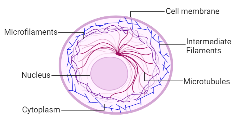
Structure of Cytoskeleton
3 Elements of Cytoskeleton
Cytoskeleton is mainly composed of three elements:
- Microfilaments
- Microtubules
- Intermediate fibers
Microfilaments
- Microfilaments are thread-like protein fibers which consist of actin monomers (f-actin).
- Each filament strand (microfilament) is composed of two helically coiled f – actin coils.
- They are 3 and 5 nm in diameter. They are normally located at the periphery of the cell and are present in bundles that together form an intracellular three – dimensional meshwork.
Microtubules
- Microtubules are small, hollow, round tubes with a diameter of about 24 nanometers.
- They consist of a single type of globular protein known as tubulin.
- Thirteen tubulins link to form a single tube.
- These help in transporting cellular materials and dividing chromosomes during cell division.
Intermediate Filaments
- Intermediate filaments are made up of a large family of polypeptides.
- They are 10 nm in diameter .
- They provide tensile strength to the cell and also facilitate the formation of keratins and neurofilaments.
Cytoskeleton – Diagram
Location of Cytoskeleton
Cytoskeleton is a protein rich filament present in the cytoplasm of all cells, including those of bacteria and archaea.
Functions of Cytoskeleton
It ensures stability, energy, and motility.
- Cytoskeleton provides shape and support to the cell
- Cytoskeleton helps in the formation of Vacuoles.
- Cytoskeleton holds different cell organelles in place.
- Cytoskeleton assists in cell signaling.
- The Cytoskeleton organizes the cell and holds the organelles of the cell in place.
- The Cytoskeleton organizes the cell and holds the organelles of the cell in place during cell division.
Significance of Cytoskeleton
These fibers in the eukaryotic cells contain a complex mesh of protein filaments and motor proteins that help in cell movement and also provides shape and support to the cell, organizes the organelles and facilitates transport of molecules, cell division and cell signaling.
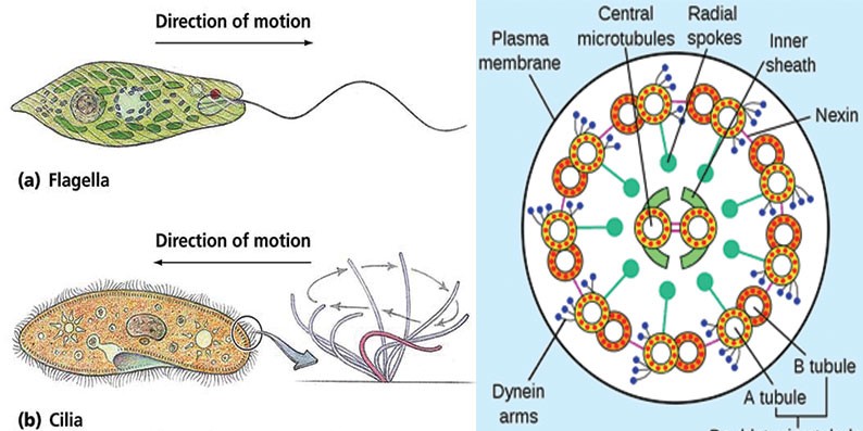
Cilia and Flagella
Cilia and Flagella – Definition
Flagella and Cilia are microscopic, contractile and filamentous processes of the cytoplasm capable of producing a current in the fluid medium for locomotion and passage of substances.
Cilia and Flagella are movable cell appendages which are structurally identical but length and function. Cilia is thicker than Flagella. Cilia are about 0.3-0.5nanometer whereas Flagella are about 0.02-0.025 nanometers thick. Flagella can be found in bacteria and sperm cells, while Cilia can be seen in species like Paramecium.
Structure of Cilia and Flagella
Cilia and Flagella are very similar in ultrastructure.
Their body parts consists of :
- Basal body
- Rootlets
- Basal plate
- Shaft.
Basal body
- The Basal body is also called kinetosome / basal granule / blepharoplast.
- The Basal body occurs embedded in the outer part of the cytoplasm below the plasma membrane.
- The Basal body is separated from the rest of the organelles by a basal plate.
- From the basal body arises Cilia and Flagella .
Rootlets
- Rootlets are striated fibrillar outgrowths that develop from the outer lower part of the basal body.
- Rootlets purpose is to provide support to the basal body.
Basal plate
- Basal plate is an area of high density which lies just above the basal body at the level of the plasma membrane.
Shaft
- It is the hair-like projecting structure of the flagellum or cilium.
- The length is 5 to 20μm in the case of the cilium and 100 to 200μm in the case of the flagellum.
- It is composed of three parts : Covering membrane (Extension of plasmalemma), Matrix, Axoneme
Location of Cilia and Flagella
Cilia are found in eukaryotic cells whereas Flagella is observed in both eukaryotic and prokaryotic cells. Cilia are present all over the surface of the cell whereas Flagella can be present on both the ends and all over the cell.
Cilia and Flagella – Diagram
Functions of Cilia and Flagella
- Cilia and Flagella serve as the locomotory organs allowing the organism to show movement.
- Cilia and Flagella create currents in the aquatic medium for obtaining food.
- Cilia and Flagella also help to capture food in some protists and animals.
- The Flagella circulate food in the gastrovascular cavity of coelenterates.
- Cilia plays an important role in the cell cycle and animal development, such as in the heart.
- Cilia also allows the internal transportation of the eggs in the oviduct and the passage of excretory substances in the kidney.
- Cilia and Flagella generate water currents for renewal of oxygen supply and quick diffusion of carbon dioxide.
- Flagella have an active role in aiding cell feeding and eukaryotic reproduction.
Significance Cilia and Flagella
- Cilia and Flagella are important structures for the purpose of movement of the cells.
- They are the means by which many microscopic unicellular and multicellular organisms move from place to place.
- Cilia and Flagella also help to move substances around cells and direct the flow of substances along tracts.
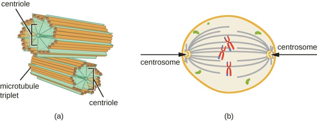
Centrosome and Centrioles
Centrosome- Definition
Centrosome is a membraneless organelle usually containing two cylindrical structures called Centrioles.
Centrioles – Definition
Centrioles are non – membranous cylindrical bodies found near the Nucleus in the Centrosome, a granular mass that serves as an organizing center for microtubules.
Structure of Centrosome and Centrioles
- The Centrosome is made up of two perpendicular Centrioles, a daughter centriole, and a mother centriole, linked together by interconnecting fibers and are positioned at right angles to each other.
- Each centriole is made of nine bundles of microtubules (three per bundle) arranged in a ring.
- The fibers of Centrioles contain tubulin, along with Lipid molecules.
- The triplet fibers are connected together by a dense material.
- An amorphous pericentriolar matrix surrounds the Centrioles. It is involved in the nucleation and anchoring of cytoplasmic microtubules.
- In post-mitotic cells, the Centrosome consists of a mature centriole and an immature centriole, known as the mother centriole and daughter centriole respectively.
Centrosome and Centrioles – Diagram
Location of Centrosome and Centrioles
- These are seen in animal cells and Flagellated plant cells but are absent in higher plants.
- The Centrosome is located in the cytoplasm usually close to the Nucleus.
Functions of Centrosome and Centrioles
- These are the centers for the organization of spindle fibers in animal cells.
- The Centrosomes help in cell division.
- Centrosomes and Centrioles maintain the chromosome number during cell division.
- Centrosomes and Centrioles become the basal bodies of Cilia and Flagella
- Centrosomes and Centrioles also bring changes in the shape of the cell membrane by phagocytosis.
Significance of Centrosomes and Centrioles
- Centrosomes and Centrioles are necessary to maintain the accurate chromosome number during cell division.
- During mitosis, it helps in organizing the microtubules ensuring that the Centrosomes are distributed to each daughter cell.
15 Comments on “Cell Organelles – The Complete Guide”