Animal Cell
Animal cell is a type of Eukaryotic cell i.e. they have a defined nucleus and membrane-bound organelles.Animal cell is the basic unit of the tissues and organs of animals.
Animal cells have a cell membrane, a Cytoskeleton, and a nucleus that contains genetic material. They also contain many organelles such as mitochondria which generates energy for the cell, Endoplasmic Reticulum and Golgi apparatus, which plays an important role in protein synthesis and transport.
Animal cells do not have a cell wall, irregular and smaller size unlike Plant Cells. They also have centrioles which play an important role in cell division. Some animal cells also have cilia and flagella which helps in cell movement. Some animal cells also have a cytoskeleton, which provides structural support and helps in cell movement.
Also Check – Plant Cell Diagram
Animal Cell Diagram
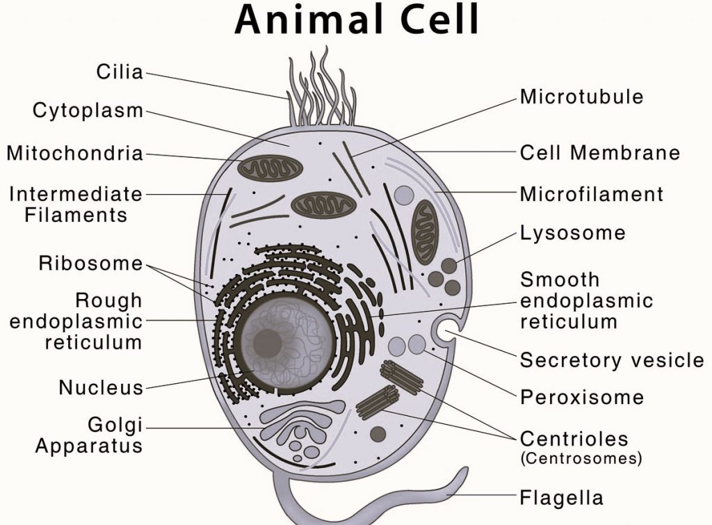
Description of Animal Cell Diagram labels is as follows
Cell Membrane
Cell membrane is a thin, flexible barrier that surrounds the Animal cell . It is called a selectively permeable Membrane as it controls the movement of materials in and out of the cell.
Also Check – Cell Organelles – The Complete Guide
Nucleus
Nucleus is the organelle of the animal cell which is in the center.It contains genetic material (DNA) and also controls the cell’s activities.
Nuclear Envelope
Nuclear envelope is the double-membrane structure that covers the nucleus .It acts as a barrier to protect the genetic material.
Nuclear Pore
Nuclear pores are openings on the nuclear envelope that allows the exchange of materials between the nucleus and the cytoplasm.
Cytoskeleton
Cytoskeleton is the network of protein fibers. It provides structure and support to the cell. It also helps to maintain cell shape and movement.
Endoplasmic Reticulum (ER)
The Endoplasmic Reticulum is the network of flattened sacs and tubules which plays an important role in protein synthesis and transport.There are two types of Endoplasmic Reticulum– ROugh Endoplasmic Reticulum and Smooth Endoplasmic Reticulum.
Also Check – 6 Important Difference between Rough and Smooth Endoplasmic Reticulum
Ribosomes
Ribosomes are small organelles which are responsible for protein synthesis.
Golgi Apparatus
Golgi apparatus is a stack of flattened sacs which plays an important role in modifying, sorting and transporting proteins and lipids.
Mitochondria
Mitochondria are the organelles that generate energy for the cell through cellular respiration. They are also called the powerhouse of the cell.
Lysosomes
Lysosomes are the small organelles that contain enzymes . It plays an important role in breakdown and recycling cellular waste.
Centrioles
Centrioles are the cylindrical small structures that are found in pairs in animal cells. Centrioles play an important role in cell division by separating the chromosomes during cell division.
Also Check – 9 Important Differences Between Plant Cell and Animal Cell
Microvilli
Microvilli are small, finger-like projections on the cell membrane. It increases the surface area of the cell .They play an important role in absorption and sensing of the cell.
Cilia
Cilia are small, hair-like structures which protrude from the surface of some cells. They play an important role in movement. Example – The movement of mucus in the respiratory tract.
Flagella
Flagella are similar to cilia. but they are longer and thicker as compared to cilia . Flagella are used for movement. For example the movement of sperm cells.
Peroxisomes
Peroxisomes are small, round cell organelles which contain enzymes that break down certain molecules like fatty acids and amino acids. They play an important role in detoxifying harmful substances in the cell.
Microfilaments
Microfilaments are thin, rod-like structures which make the cytoskeleton of the animal cell. They provide structural support and help to maintain the shape of the animal cell.
Intermediate filaments
Intermediate filaments are thicker and stronger than microfilaments.it also makes up the cytoskeleton. They also provide structural support to the cell and help to resist mechanical stress.
Did you find this article helpful? We’d love to hear your thoughts and suggestions in the comments!
Also Check – 16 Important Differences between Prokaryotic and Eukaryotic cell
Also Check – What is the Difference between Plant Vacuoles and Animal Vacuoles?
Also Check – 10 Important Difference between Cell organelles and Cell Inclusions
2 Comments on “Animal Cell Diagram”