Endocytosis is a fundamental process that occurs within cells, enabling them to internalise substances from their external environment. It is a vital mechanism by which cells obtain the necessary nutrients for growth and development. This process involves the uptake of various substances, including fluids, electrolytes, proteins and other macromolecules and the subsequent breakdown of these substances into smaller elements for either use by the cell or elimination purposes.
Table of Contents
Definition of Endocytosis
Endocytosis is a cellular process that involves the uptake of materials from the external environment into the cell. The process involves the formation of a vesicle or sac-like structure around the material, which then fuses with the cell’s membrane, allowing the material to be taken into the cell.
One of the critical roles of Endocytosis is in the immune system, where white blood cells utilise this mechanism to capture and destroy potential Pathogens, including Bacteria and Protists. They entrap the Pathogens and break them down for elimination from the body.
Discovery
Endocytosis was first described by Christian de Duve, a Belgian cytologist and biochemist who won several Nobel prizes for his role in discovering cellular elements such as lysosomes, peroxisomes, endosomes and even exocytosis cellular mechanisms, including the Endocytosis mechanism. In this article, we will delve deeper into the process of Endocytosis, its different types and its significance in cellular biology.
Also Check – Differences and Differences Between Endocytosis and Exocytosis
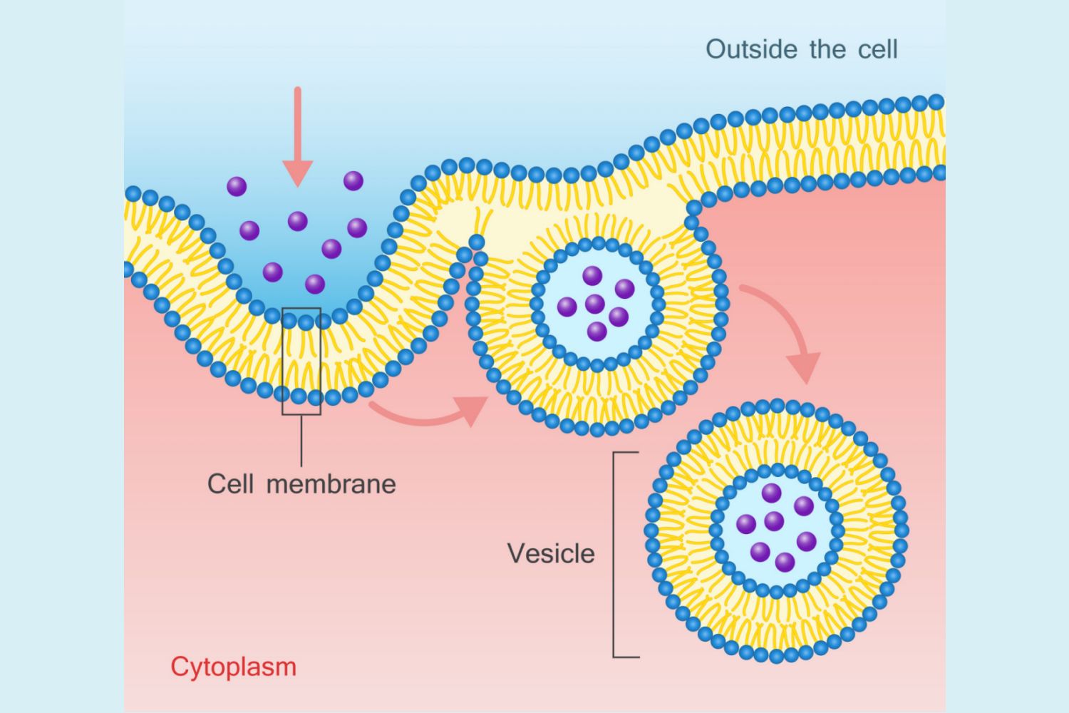
Mechanism of Endocytosis
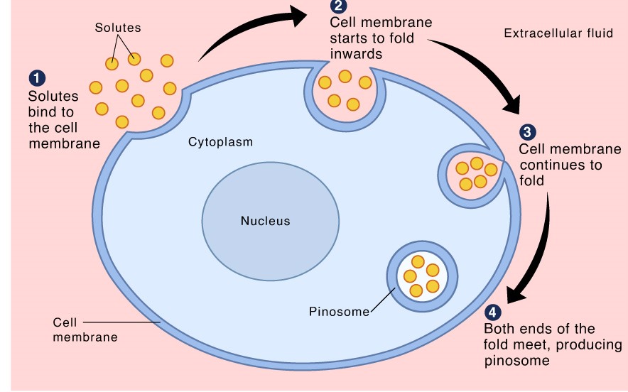
3 Steps of Endocytosis
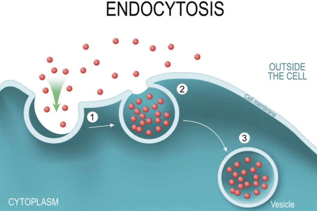
- Invagination
The plasma membrane folds inward, forming a cavity that fills with extracellular fluid, dissolved molecules, food particles, foreign matter, Pathogens, or other substances. This process is known as invagination.
- Vesicle formation
The plasma membrane then folds back on itself until the ends of the in-folded membrane meet, trapping the fluid inside The Vesicle. In some cells, long channels also form extending from the membrane deep into the cytoplasm. This enclosed membrane or cavity is known as a vesicle.
- Vesicle detachment
The Vesicle is then pinched off from the membrane as the ends of the in-folded membrane fuse together. The internalised vesicle is then processed by the cell.
These three steps of Endocytosis are crucial for cells to internalise substances from their external environment and play an essential role in cellular biology.
Role of Cell Membrane in Endocytosis
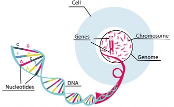
The Cell Membrane, also known as the plasma membrane, plays a critical role in Endocytosis. For Endocytosis to occur, substances must be enclosed within a vesicle formed from The Cell Membrane. The main components of The Cell Membrane are proteins and lipids that aid in Cell Membrane flexibility and molecule transport.
Phospholipids are the primary lipid molecules in The Cell Membrane that form a double-layered barrier between the external cellular environment and the cell’s interior. These lipids have hydrophilic (attracted to water) heads and hydrophobic (repelled by water) tails. In contact with liquid, they spontaneously arrange so that their hydrophilic heads face the cytosol and extracellular fluid, while their hydrophobic tails move away from the fluid to the internal region of the lipid bilayer membrane.
The Cell Membrane is an essential component of the Endocytosis process and its ability to form vesicles is critical for internalising substances from the external environment.
The Cell Membrane is semi-permeable that means only certain molecules can diffuse across the membrane. Substances that cannot diffuse across The Cell Membrane must be helped across by passive diffusion processes (facilitated diffusion), active transport (which requires energy), or by Endocytosis. Endocytosis involves the removal of portions of The Cell Membrane for the formation of vesicles and internalisation of substances.
In addition to its role in Endocytosis The Cell Membrane also plays a crucial role in maintaining cell size. Membrane components must be replaced periodically and this is accomplished by the process of Exocytosis.
Exocytosis involves the formation, transportation and fusion of internal vesicles with The Cell Membrane to expel substances from the cell.
3 Types Of Endocytosis
There are 3 types of Endocytosis –
- Phagocytosis
- Pinocytosis
- Receptor-mediated Endocytosis
Phagocytosis
Definition
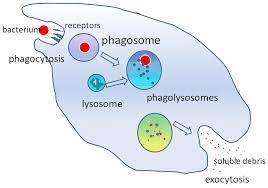
Phagocytosis is a process by which cells engulf and enclose particles within folded membranes to form a Phagosome. Phagosomes are later processed by cellular enzymes. This mechanism is commonly used by white blood cells such as the macrophages, monocytes, neutrophils, eosinophils and dendritic cells, for eliminating Pathogens from the system. Some protozoans, such as Entamoeba use Phagocytosis to acquire nutrients.
The mechanism of Phagocytosis was first observed by Canadian physician William Osler in 1876.
5 Steps of Phagocytosis
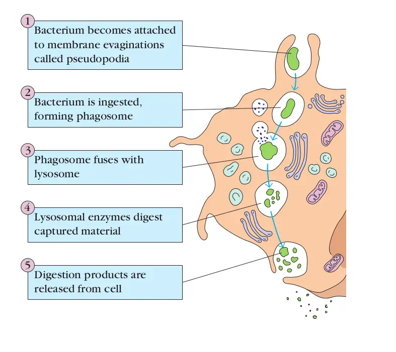
Phagocytosis is carried out in five basic steps –
- Detection
The Phagocytic cell detects the molecule of interest or an antigen and moves towards it.
- Attachment
The Phagocyte attaches itself to the target molecule or antigen. Phagocytes can extend their membrane, called pseudopodia, to surround the particle or Pathogen. The pseudopodia extend towards each other while enclosing the particles.
- Ingestion
The particle is enclosed within a vesicle formed from the extended pseudopodia that have fused. This vesicle with the enclosed particles is known as a Phagosome.
- Fusion
The Phagosome fuses with the lysosomes of the Phagocyte forming a Phagolysosome. The lysosomes contain digestive enzymes that degrade or digest the materials contained in The Vesicle.
Also Check – Lysosomes – Class 9th Definition , Functions , Location and Importance of Lysosomes
- Elimination
The degraded particles are then expelled from the Phagocytic cell by exocytosis.
Phagocytosis in Immune Cells
In immune cells, Phagocytosis helps to eliminate Bacteria, cancer cells, virus-infected cells, or other harmful substances. It is carried out by specialised immune cells, such as macrophages.
The Basic Steps of Phagocytosis in Immune Cells
The process of Phagocytosis in immune cells involves the following steps –
- Detection- The Phagocyte detects the antigen, such as a bacterium and moves towards the target cell.
- Attachment- The Phagocyte makes contact with and attaches to the bacterium. This binding initiates the formation of pseudopodia that surround the bacterium.
- Ingestion- The surrounded bacterium is enclosed within a vesicle formed when pseudopodia membranes fuse. This vesicle with bacterium enclosed, called a Phagosome, is internalized by the Phagocyte.
- Fusion- The Phagosome fuses with an organelle called a lysosome and becomes known as a Phagolysosome. Lysosomes contain enzymes that digest organic material. The release of digestive enzymes within the Phagolysosome degrades the bacterium.
- Elimination- The degraded material is expelled from the cell by exocytosis.
Also Check – How Amoeba Acquires its Food through the Process of Phagocytosis
Phagocytosis in Protists
In Protists, Phagocytosis is the primary means by which these organisms obtain food. It occurs similarly to Phagocytosis in immune cells, It is more commonly used for acquiring nutrients.
Also Check – Nutrition in Amoeba
Also Check – Nutrition in Paramecium
Pinocytosis
Definition
Pinocytosis is a type of Endocytosis where small particles in extracellular fluids enter the cell through The Cell Membrane by invagination, forming a small vesicle with suspended small molecules or particles within a cell. Pinocytosis is also known as cell drinking or fluid Endocytosis.
Mechanism of Pinocytosis
Pinocytosis works similarly to other endocytic processes. The major difference between Pinocytosis and Phagocytosis is that in Pinocytosis, the particles ingested are contained within the cell’s extracellular fluids. The Cell Membrane invaginates together with The Vesicle containing the fluid particles, transporting them into the cell lysosomes. The Vesicle and the lysosomes fuse, releasing digestive enzymes from the lysosomes. The enzymes degrade The Vesicle, releasing its content into the cell cytoplasm for utilisation by the cell. Sometimes, The Vesicles do not interact with the lysosomes but instead move across the cell, fusing with The Cell Membrane, causing a recycling effect of the membrane proteins and lipids.
Types of Pinocytosis
Pinocytosis takes place by two mechanisms-
MicroPinocytosis
The formation of small vesicles of about 0.1 um diameter. It takes place in the body cells, forming tiny budding vesicles on The Cell Membrane known as caveolae. They are found in the blood vessel endothelium.
MacroPinocytosis
The formation of larger vesicles of about 0.5-5um in diameter. They are found on the white blood cells. The large vesicles are formed by The Cell Membrane ruffles (villi), which are projections that extend to the extracellular fluids and have the ability to fold back by themselves. While folding, they shovel in some of the extracellular fluid forming a vesicle that pulls into the cell.
Receptor-Mediated Endocytosis
Definition
Receptor-mediated Endocytosis is a type of Endocytosis that is used by cells to selectively internalise specific molecules. This process requires the binding of the molecule to specific receptors on The Cell Membrane before it is internalised by Endocytosis. This method of Endocytosis is highly specific and it allows cells to regulate the type and amount of molecules that are taken up.
Membrane Receptors and Clathrin-coated Pits
Membrane receptors are proteins that are found on the surface of The Cell Membrane. They are involved in various cellular processes, including signal transduction, cell adhesion and Endocytosis.
In receptor-mediated Endocytosis, these receptors are found in regions of the plasma membrane that are coated with a protein called clathrin. These regions are known as Clathrin-coated pits.
Basic Steps of Receptor-mediated Endocytosis
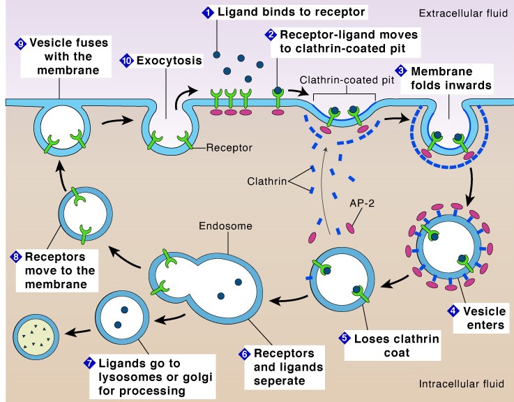
- Binding of the specified molecule to the receptor on the plasma membrane.
- Migration of the molecule-bound receptor along the membrane to a region containing a clathrin-coated pit.
- Accumulation of molecule-receptor complexes in the clathrin-coated pit that forms an invagination that is internalised by Endocytosis.
- Formation of a clathrin-coated vesicle that encapsulates the ligand-receptor complex and extracellular fluid.
- Fusion of the clathrin-coated vesicle with an endosome in the cytoplasm and removal of the clathrin coating.
- Enclosure of the receptor in a lipid membrane for recycling back to the plasma membrane or degradation of the specified molecule by lysosomal enzymes.
After the molecule binds to the receptor, the pit regions are internalised and clathrin-coated vesicles are formed. These vesicles contain the ligand-receptor complex and extracellular fluid. Once they fuse with early endosomes, the clathrin coating is removed from The Vesicles and the contents are emptied into the cell.
Receptor-mediated Endocytosis is thought to be more than a hundred times more efficient at taking in selective molecules than Pinocytosis. This is because receptor-mediated Endocytosis is a highly regulated process that requires specific interactions between the ligand and receptor, while Pinocytosis is a non-specific process that involves the uptake of extracellular fluid and random particles.
Examples of Endocytosis
Examples of Phagocytosis
Immune cells such as macrophages, dendritic cells and neutrophils use Phagocytosis to engulf and digest particles like Bacteria, fungi, dust particles and dead cells. Macrophages, the largest Phagocytic cells in the immune system, detect and attach to antigens before ingesting and digesting them. The ingested particles are released from the cell by exocytosis.
Examples of Pinocytosis-
Pinocytosis is the uptake or absorption of nutrients in the small intestines.
Examples of Receptor-Mediated Endocytosis
Iron-bound transferrin recycling , Receptor-mediated Endocytosis – This is the uptake of cholesterol bound to low-density lipoprotein (LDL), a complex of phospholipid, protein and cholesterol.
Frequently asked questions on Endocytosis
Question- What is Endocytosis?
Answer- Endocytosis is a cellular process that involves the uptake of materials from the external environment into the cell by forming a vesicle or sac-like structure around the material, which then fuses with the cell’s membrane, allowing the material to be taken into the cell.
Question- What are the steps of Endocytosis?
Answer- The three steps of Endocytosis are invagination, vesicle formation and vesicle detachment.
Question-How does The Cell Membrane play a role in Endocytosis?
Answer- The Cell Membrane plays a crucial role in Endocytosis as it is responsible for forming vesicles that internalise substances from the external environment.
Question-What is Phagocytosis?
Answer- Phagocytosis is a cellular process where cells engulf and enclose particles within folded membranes to form a Phagosome, which is later processed by cellular enzymes.
Question- What are the five steps of Phagocytosis?
Answer- The five steps of Phagocytosis are detection, attachment, engulfment, Phagosome formation and digestion.
Question- What is Phagocytosis and how is it used in Protists?
Answer- Phagocytosis is a process that allows cells to engulf and enclose particles within a folded membrane to form a Phagosome. In Protists, Phagocytosis is the primary means by which these organisms obtain food. It occurs similarly to Phagocytosis in immune cells, but it is more commonly used for acquiring nutrients.
Question- What are the five basic steps involved in Phagocytosis?
Answer- The five basic steps involved in Phagocytosis are detection, attachment, ingestion, fusion and elimination.
Question- What is Pinocytosis and how does it work?
Answer- Pinocytosis is a type of Endocytosis where small particles in extracellular fluids enter the cell through The Cell Membrane by invagination, forming a small vesicle with suspended small molecules or particles within a cell. Pinocytosis works similarly to other endocytic processes and the major difference between Pinocytosis and Phagocytosis is that in Pinocytosis, the particles ingested are contained within the cell’s extracellular fluids.
Question- What are the two mechanisms by which Pinocytosis takes place?
Answer- Pinocytosis takes place by two mechanisms- MicroPinocytosis and MacroPinocytosis. MicroPinocytosis involves the formation of small vesicles of about 0.1 um diameter and it takes place in the body cells, forming tiny budding vesicles on The Cell Membrane known as caveolae. MacroPinocytosis involves the formation of larger vesicles of about 0.5-5 um in diameter and they are found on the white blood cells.
Question- How is receptor-mediated Endocytosis different from other types of Endocytosis?
Answer- Receptor-mediated Endocytosis is a type of Endocytosis that is used by cells to selectively internalise specific molecules. This process requires the binding of the molecule to specific receptors on The Cell Membrane before it is internalised by Endocytosis. This method of Endocytosis is highly specific and it allows cells to regulate the type and amount of molecules that are taken up.
Question- What are membrane receptors and how are they involved in receptor-mediated Endocytosis?
Answer- Membrane receptors are proteins that are found on the surface of The Cell Membrane. They are involved in various cellular processes, including signal transduction, cell adhesion and Endocytosis. In receptor-mediated Endocytosis, these receptors are found in regions of the plasma membrane that are coated with a protein called clathrin.
Question- What are the basic steps of receptor-mediated Endocytosis?
Answer- The basic steps of receptor-mediated Endocytosis include binding of the specified molecule to the receptor on the plasma membrane, migration of the molecule-bound receptor along the membrane to a region containing a clathrin-coated pit, accumulation of molecule-receptor complexes in the clathrin-coated pit, formation of a clathrin-coated vesicle that encapsulates the ligand-receptor complex and extracellular fluid, fusion of the clathrin-coated vesicle with an endosome in the cytoplasm and removal of the clathrin coating and enclosure of the receptor in a lipid membrane for recycling back to the plasma membrane or degradation of the specified molecule by lysosomal enzymes.
5 Comments on “Endocytosis -Definition, Mechanism, Types, Examples”