Human Heart
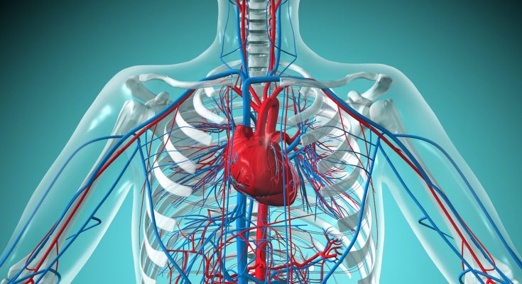
The Human Heart is a muscular structure responsible for the Circulation of Blood throughout the body. Along with the Blood Vessels, it forms the Cardiovascular system. The Heart is a remarkable organ that plays a vital role in keeping us alive by delivering Oxygen and essential nutrients to every part of the body through the pumping of Blood.
Definition
The Human Heart is a vital organ that pumps blood throughout the body. It is a muscular organ located in the chest, behind the breastbone, and between the lungs. The heart has four chambers – Two upper chambers called Atria and Two lower chambers called Ventricles. Its main function is to circulate blood, which delivers oxygen and nutrients to the body’s tissues and organs while removing waste products. The heart works by contracting and relaxing in a rhythmic manner, controlled by electrical signals.
Table of Contents
Location of Human Heart in the Body
The Human Heart is located between the lungs in the middle of the chest, behind and slightly to the left of the breastbone (sternum). A double-layered membrane called the Pericardium surrounds the Heart like a sac. The outer layer of the Pericardium surrounds the roots of the Heart’s major Blood Vessels and is attached by ligaments to the spinal column, diaphragm and other parts of the body.
The Heart is positioned in the Mediastinum which is the central compartment of the thoracic cavity. It’s important to note that the exact location of the Heart can vary slightly from person to person, depending on factors such as age, gender, body size and overall health.
Structure of the Heart
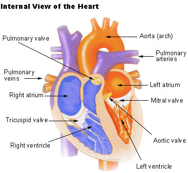
The Human Heart is a complex organ that is divided into four chambers, each with its unique purpose. The Right Atrium and Ventricle make up the “right Heart,” while The Left Atrium and Ventricle make up the “left Heart.” The structure of the Heart also includes the Aorta, the largest artery in the body that carries Oxygenated Blood from the Heart to the rest of the body.
ALso Check – Neatly Labelled Easy to Draw Human Heart Diagram
External Structure of the Heart
The external structure of the Heart is the part that can be seen without any dissection. One of the first structures observed is the Pericardium. The Human Heart is situated on the left side of the chest and is enclosed within a fluid-filled cavity called the pericardial cavity. The walls and lining of the pericardial cavity are made up of a membrane known as the Pericardium.
Pericardium
The Pericardium is a fibrous membrane found as an external covering around the Heart. It serves to protect the Heart by producing a serous fluid that lubricates the Heart and prevents friction between surrounding organs. The Pericardium also helps by holding the Heart in its position and maintaining a hollow space for the Heart to expand itself when it is full. The Pericardium has two exclusive layers-
- The Visceral Layer- it directly covers the outside of the Heart.
- Parietal Layer – It forms a sac around the outer region of the Heart that contains the fluid in the pericardial cavity.
Structure of the Heart Wall
The Heart wall is made up of three layers
- Epicardium
- Myocardium
- Endocardium
Epicardium
The Epicardium is the outermost layer of the Heart. It is composed of a thin-layered membrane that serves to lubricate and protect the outer section.
Myocardium
This layer of muscle tissue constitutes the middle layer wall of the Heart. It contributes to the thickness of the Heart and is responsible for the pumping action.
Endocardium
The Endocardium is the innermost layer that lines the inner Heart chambers and covers the Heart Valves. It is responsible for preventing the Blood from sticking to the inner walls, thereby preventing potentially fatal Blood clots.
Also Check – The Human Heart- A Guide for Middle School Students
Internal Structure of Heart
The Human Heart has a complex internal structure with several chambers and Valves that control the flow of Blood. The internal structure of the Heart can be divided into four chambers, namely, The Left Atrium, Right Atrium, Left Ventricle and Right Ventricle. The Atria are thin-walled and smaller than the Ventricles. They are responsible for receiving Blood from the large veins, while the Ventricles are larger and more muscular chambers responsible for pumping and pushing Blood out into Circulation.
4 Chambers of the Heart
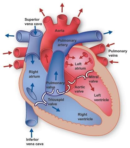
The Heart is a muscular organ that consists of four chambers, two Atria and two Ventricles. The chambers of the Heart can be classified based on the number of chambers present in the vertebrate Heart. In Humans, being mammals, we have four chambers-
- The Left Atrium
- The Right Atrium
- The Left Ventricle
- The Right Ventricle.
Atria
The Atria are thin-walled chambers that have less muscular walls compared to the Ventricles. They are smaller in size and are the Blood-receiving chambers that are fed by the large veins. The Right Atrium receives deOxygenated Blood from the body through the Superior and Inferior Vena Cava, while The Left Atrium receives Oxygenated Blood from the lungs through the pulmonary veins.
Ventricles
The Ventricles are larger and more muscular chambers that are responsible for pumping and pushing Blood out into Circulation. They are connected to larger arteries that deliver Blood for Circulation throughout the body. The Left Ventricle is the largest and most muscular of all the chambers and it is responsible for pumping Oxygenated Blood to the entire body. The Right Ventricle, on the other hand, is comparatively smaller than the Left Ventricle and is responsible for pumping deOxygenated Blood to the lungs for Oxygenation.
Function of the Chambers
The Right Atrium and Right Ventricle are smaller than the left chambers and the walls consist of fewer muscles compared to the left portion. The Blood originating from the right side flows through the pulmonary Circulation, while Blood arising from the left chambers is pumped throughout the body. The Left Atrium and Left Ventricle, being the largest chambers, have thicker walls due to the higher pressure required to pump Oxygenated Blood to the entire body.
Blood Vessels
Blood Vessels are an essential part of the circulatory system in all vertebrates, including Humans. The Vessels transport Blood throughout the body and can be classified into three types- veins, capillaries and arteries.
Veins-
- Veins are Blood Vessels that carry deOxygenated Blood from the body back to the Heart. The Superior and Inferior Vena Cava are the largest veins in the body and deliver Blood into the Right Atrium of the Heart. Veins typically have thin walls and low Blood pressure, but have Valves to prevent backflow of Blood.
Capillaries-
- Capillaries are the smallest type of Blood Vessel and form a network that connects arterioles (small arteries) to venules (small veins). They are responsible for exchanging Oxygen and nutrients with tissues and removing waste products such as carbon dioxide. Capillaries have very thin walls, allowing for the exchange of gases and other molecules.
Arteries-
- Arteries are muscular-walled tubes that carry Oxygenated Blood away from the Heart to all parts of the body. The largest artery in the body is the Aorta. Aorta branches off into smaller arteries that supply Blood to specific regions of the body. Arteries have thick walls and high Blood pressure to withstand the force of Blood being pumped out of the Heart.
Also Check – Arteries in The Body
The Blood Vessels in the Heart also play a crucial role in maintaining Blood flow through the organ. The Coronary arteries supply Oxygen and nutrients to the Heart muscle, while the cardiac veins remove waste products. The Blood Vessels in the Heart are arranged in a network, with the Coronary arteries branching off the Aorta and the cardiac veins draining into the Right Atrium.
Also Check – 8 Difference Between Arteries , Veins and Capillaries
Valves in the Heart
The Heart contains flaps of fibrous tissue called Valves, which are crucial for maintaining Blood flow in a single direction and preventing backflow.
There are two types of Valves in the Heart
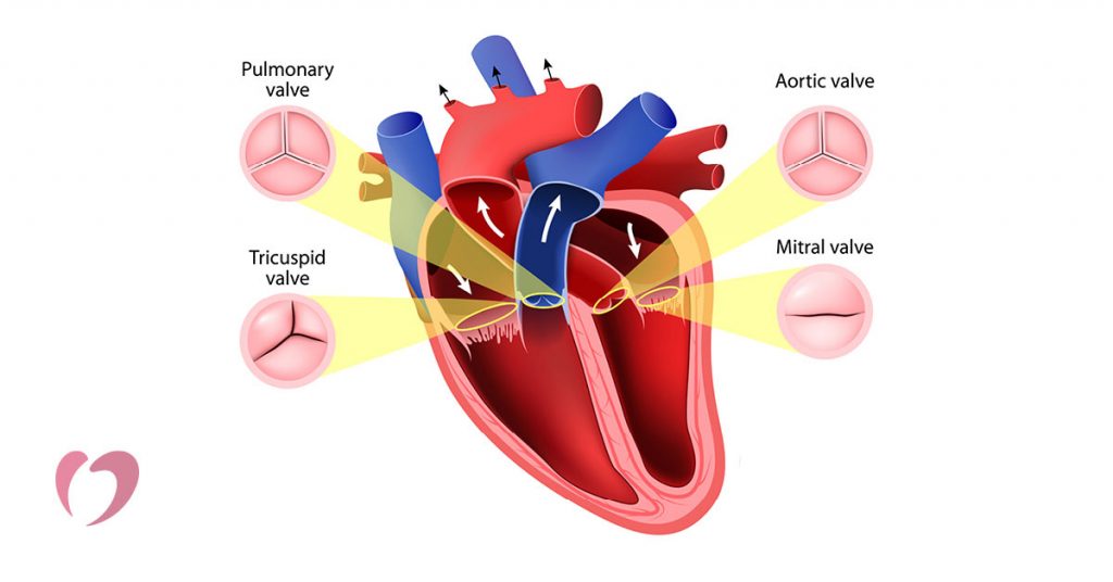
- Atrioventricular Valves
- Semilunar Valves
Atrioventricular Valves
Atrioventricular Valves are located between the Ventricles and Atria. They include –
- The Tricuspid Valve – It is found between the Right Ventricle and Right Atrium .
- The Mitral Valve. It is located between the Left Ventricle and left Atrium.
These Valves open to allow Blood to flow from the Atria into the Ventricles and then close to prevent backflow of Blood into the Atria during ventricular contraction.
Semilunar Valves
Semilunar Valves are located between the Left Ventricle and the Aorta and between the pulmonary artery and the Right Ventricle. These Valves have three cusps that open during ventricular contraction to allow Blood to flow into the arteries and then close to prevent backflow of Blood into the Ventricles during relaxation.
Also Check – Heart Valves- Types, Location, Structure and Functions
Circulation- The Process of Blood Flow in the Body
Blood Circulation is the process by which Blood flows through the body, supplying vital organs and tissues with Oxygen and nutrients while removing waste products. The circulatory system is made up of the Heart, Blood Vessels and Blood.
Types of Circulation
There are three main types of Circulation in the Human body-
- Pulmonary Circulation
- Systemic Circulation
- Coronary Circulation
Pulmonary Circulation
- Pulmonary Circulation is responsible for carrying deOxygenated Blood away from the Heart to the lungs.
- In Lungs it is Oxygenated and then brings the Oxygenated Blood back to the Heart. This process is essential for the body to receive Oxygen.
Systemic Circulation
- Systemic Circulation is responsible for the Oxygenated Blood being pumped from the Heart to every organ and tissue in the body.
- The deOxygenated Blood then returns to the Heart to be pumped to the lungs for Oxygenation. This process is vital for the functioning of all organs in the body.
Also Check – Double Circulation -Definition, 2 Loops, Flowchart,Types, Importance
Coronary Circulation
- The Heart itself is a muscle and requires a constant supply of Oxygenated Blood. This is where Coronary Circulation comes into play.
- It is the Circulation responsible for supplying Oxygenated Blood to the Heart.
- Without proper Coronary Circulation, the Heart cannot function properly and can lead to Heart diseases.
The Role of the Heart in Circulation
- The Heart is a vital organ in the circulatory system and its main function is to pump Blood throughout the body. It consists of four chambers- two Atria and two Ventricles. The Atria are the upper chambers that receive Blood, while the Ventricles are the lower chambers that pump Blood out of the Heart.
- The right side of the Heart receives deOxygenated Blood from the body and pumps it to the lungs for Oxygenation through the pulmonary artery. The Oxygenated Blood then returns to the left side of the Heart through the pulmonary veins.
- The left side of the Heart receives the Oxygenated Blood from the lungs and pumps it to the rest of the body through the Aorta. This process ensures that every cell in the body receives the Oxygen and nutrients it needs to function correctly.
Blood Vessels Entering and Leaving The Heart
The Heart is the central organ of the circulatory system that pumps Blood to all parts of the body. The Blood Vessels that enter and leave the Heart play a crucial role in this process.
Blood Vessels Entering The Heart
The right auricle receives Blood from two large Vessels
- The Superior Vena Cava/Anterior Vena Cava
- The Inferior Vena Cava/Posterior Vena Cava.
Superior Vena Cava/Anterior Vena Cava
The Superior Vena Cava/Anterior Vena Cava brings deOxygenated Blood from the upper regions of the body, including the head, chest and arms.
The Inferior Vena Cava/Posterior Vena Cava
The Inferior Vena Cava/Posterior Vena Cava brings Blood from the lower region of the body, including the abdomen and legs.
The left Atrium receives four pulmonary veins, two from each lung. The pulmonary veins bring Oxygenated Blood, which is an essential part of the circulatory system.
Blood Vessels Leaving the Heart-
Blood Vessels leaving the Heart arise from the Ventricles and are two large Blood Vessels.
- Pulmonary Artery
- Aorta
Pulmonary Artery
The pulmonary artery arises from the Right Ventricle and carries deOxygenated Blood to the lungs for Oxygenation. The pulmonary artery splits into two branches, one for each lung and further divides into smaller arterioles and capillaries within the lungs. In the lungs, the Blood is enriched with Oxygen and releases carbon dioxide, a waste product, which is expelled from the body during exhalation.
Aorta
The Aorta is the largest artery in the body and arises from the Left Ventricle. It carries Oxygenated Blood to supply it to all parts of the body. The Aorta is divided into different sections, including the ascending Aorta, the aortic arch and the descending Aorta. The ascending Aorta carries Blood from the Left Ventricle and then turns into the aortic arch, which curves and branches into the brachiocephalic artery, the left common carotid artery and the left subclavian artery. These branches supply Blood to the head, neck and arms. The descending Aorta carries Blood to the rest of the body, including the abdomen and legs.
Role of Heart Valves
To ensure that Blood flows in one direction only, the Heart has four Valves- The Tricuspid Valve, the pulmonary Valve, The Mitral Valve and the aortic Valve. The Tricuspid Valve is situated between the Right Atrium and the Right Ventricle, while the pulmonary Valve is located between the Right Ventricle and the pulmonary artery. The Mitral Valve is situated between The Left Atrium and the Left Ventricle and the aortic Valve is located between the Left Ventricle and the Aorta. These Valves open and close in a specific sequence to allow Blood flow and prevent backward flow or regurgitation of Blood.
Pumping Action of the Heart – How the Human Heart Works
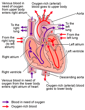
- The Heart is a muscular organ that pumps Blood throughout the body. It is divided into four chambers – two upper chambers called Atria and two lower chambers called Ventricles. The Heart’s pumping action is controlled by electrical signals that travel through specialised cells in the Heart.
- The Right Atrium receives deOxygenated Blood from the body through the Superior and Inferior Vena Cava. When the Right Atrium expands, Blood flows into the chamber and then contracts, forcing the Blood into the Right Ventricle.
- The Right Ventricle then contracts and the Blood is pumped through the pulmonary artery to the lungs, where it gets Oxygenated. The Oxygenated Blood then returns to the Heart through the pulmonary veins, which enter The Left Atrium. During this time, The Left Atrium is relaxed.
- Once The Left Atrium has collected Oxygenated Blood, it contracts, forcing the Blood into the Left Ventricle. At the same time, the Right Ventricle expands to receive deOxygenated Blood from the Right Atrium.
- The Left Ventricle then contracts, pumping the Oxygenated Blood through the Aorta, which carries it to all parts of the body. The Right Ventricle contracts and pumps the deOxygenated Blood to the lungs, completing the cycle.
- Valves play a crucial role in ensuring that Blood flows in a single direction, preventing backflow. There are two types of Valves – Atrioventricular Valves and Semilunar Valves. Atrioventricular Valves are located between the Atria and Ventricles and Semilunar Valves are located between the Ventricles and the arteries.
Also Check – How Blood Flow in Heart ?
Function of the Heart in Human body
The Heart is a muscular organ that plays a vital role in the circulatory system of the Human body. It is responsible for pumping Blood throughout the body, delivering Oxygen and nutrients to the tissues and organs and removing waste products.
The primary functions of the Heart are as follows-
- Circulating Blood- The Heart is responsible for circulating Blood throughout the body, which is essential for the transport of Oxygen, nutrients, hormones and other substances to the tissues and organs. The Blood also carries waste products such as carbon dioxide away from the tissues and organs, which are then eliminated from the body.
- Maintaining Blood Pressure- The Heart helps to maintain Blood pressure within the circulatory system. It does this by pumping Blood with enough force to ensure that it flows through the arteries and capillaries, which are the smaller Blood Vessels and returns to the Heart via the veins.
- Regulation of Blood Flow- The Heart plays a critical role in regulating Blood flow to different parts of the body. It can increase or decrease Blood flow to different organs and tissues, depending on their needs. For example, during exercise, the Heart pumps more Blood to the muscles to provide them with Oxygen and nutrients.
- Oxygenation of Blood- The Heart is responsible for Oxygenating the Blood. It receives deOxygenated Blood from the body and pumps it to the lungs, where it picks up Oxygen and releases carbon dioxide. The Oxygenated Blood then returns to the Heart and is pumped out to the rest of the body.
- Removal of Waste Products- The Heart also helps to remove waste products from the body. It receives Blood that has been depleted of Oxygen and nutrients and pumps it to the lungs and other organs, where the waste products are removed from the body.
Also Check – 13 Important Functions of Heart
Heartbeat
Heartbeats are a crucial aspect of the Cardiovascular system. They represent the synchronised contraction and relaxation of the Heart muscles, which is initiated by an electrical impulse generated in the sinoAtrial (SA) node.
The Electrical Conduction System of the Heart
- The electrical conduction system of the Heart consists of specialised cardiac muscle cells that have an intrinsic ability to generate and conduct electrical impulses.
- The SA node, located in the Right Atrium, generates the electrical impulse that spreads through the Atria, causing them to contract.
- The impulse then reaches the Atrioventricular (AV) node, where it is briefly delayed before continuing down the bundle of His and the Purkinje fibres that are responsible for stimulating ventricular contraction.
Process of Heartbeat
- The process of Heartbeat begins with the contraction of the Atria that occurs when the electrical impulse from the SA node reaches them.
- This contraction forces Blood from the Atria into the Ventricles through the AV Valves, which are open at this point.
- After a brief delay at the AV node, the electrical impulse reaches the Ventricles, causing them to contract and forcing the Blood out of the Heart through the Semilunar Valves and into the circulatory system.
Importance of Heart Rate and Rhythm
The Heart rate and rhythm are critical in maintaining proper Blood flow and Oxygenation to the body. A healthy Heart rate ranges between 60-100 beats per minute and an irregular Heart rhythm, such as Atrial fibrillation, can result in poor Blood flow, Blood clots and other complications. In contrast, regular exercise and a healthy lifestyle can contribute to a lower resting Heart rate and better overall Heart health.
Blood Pressure
Blood pressure is the force that Blood exerts on the walls of arteries. It is expressed in two numbers, representing the upper (systolic) and lower (diastolic) limits of pressure.
- Systolic Pressure- This is the pressure when fresh Blood is pushing through the artery as a result of ventricular contraction of the Heart.
- Diastolic Pressure- This is the pressure recorded when the wave has passed over.
Also Check – Why Do Arteries Have Thick Elastic Walls ?
Normal Blood Pressure
The normal Blood pressure range for adults is-
- Systolic- 100-140 mm
- Diastolic- 60-80 mm
High Blood Pressure
A Blood pressure reading above 140/90 is considered hypertension (high Blood pressure). Hypertension can increase the risk of Heart disease, stroke and other health problems.
Also Check – What’s Systolic and Diastolic Blood Pressure?
Measuring Blood Pressure
- Blood pressure can be measured using an instrument called a sphygmomanometer. This device consists of an inflatable cuff that is wrapped around the upper arm and a pressure gauge.
- During a Blood pressure measurement, the cuff is inflated to temporarily cut off Blood flow to the artery. The gauge then measures the pressure when the cuff is slowly released. This gives a reading of both the systolic and diastolic pressure.
Frequently Asked Questions Answers on Human Heart
What is the role of the Human Heart in the body?
Answer- The Human Heart is responsible for the Circulation of Blood throughout the body, delivering Oxygen and essential nutrients to every part of the body through the pumping of Blood.
What is the structure of the Heart and where is it located in the body?
Answer-The Human Heart is a complex organ that is divided into four chambers and is located between the lungs in the middle of the chest, behind and slightly to the left of the breastbone (sternum). It is surrounded by a double-layered membrane called the Pericardium.
What is the Pericardium and what is its function?
Answer- The Pericardium is a fibrous membrane found as an external covering around the Heart. It serves to protect the Heart by producing a serous fluid that lubricates the Heart and prevents friction between surrounding organs. The Pericardium also helps by holding the Heart in its position and maintaining a hollow space for the Heart to expand itself when it is full.
What are the different layers that make up the Heart wall?
Answer- The Heart wall is made up of three layers, namely the Epicardium, Myocardium and Endocardium. The Epicardium is the outermost layer, the Myocardium is the middle layer responsible for the pumping action and the Endocardium is the innermost layer that lines the inner Heart chambers and covers the Heart Valves.
What are the chambers of the Heart and what are their functions?
Answer-The Heart consists of four chambers, two Atria and two Ventricles. The Atria are responsible for receiving Blood from the large veins, while the Ventricles are larger and more muscular chambers responsible for pumping and pushing Blood out into Circulation. The Right Atrium receives deOxygenated Blood from the body, while The Left Atrium receives Oxygenated Blood from the lungs. The Right Ventricle pumps deOxygenated Blood to the lungs for Oxygenation, while the Left Ventricle pumps Oxygenated Blood to the entire body.
What are the two types of Valves found in the Heart?
Answer- The two types of Valves found in the Heart are Atrioventricular Valves and Semilunar Valves.
Where are the Atrioventricular Valves located and what are their functions?
Answer- The Atrioventricular Valves are located between the Ventricles and Atria. Their function is to allow Blood to flow from the Atria into the Ventricles and then close to prevent backflow of Blood into the Atria during ventricular contraction.
What are the Semilunar Valves and where are they located?
Answer- Semilunar Valves are located between the Left Ventricle and the Aorta and between the pulmonary artery and the Right Ventricle. They have three cusps that open during ventricular contraction to allow Blood to flow into the arteries and then close to prevent backflow of Blood into the Ventricles during relaxation.
What are the primary functions of the Heart in the Human body?
Answer- The primary functions of the Heart are circulating Blood, maintaining Blood pressure, regulating Blood flow, Oxygenating the Blood and removing waste products.
What are the three main types of Circulation in the Human body?
Answer- The three main types of Circulation in the Human body are pulmonary Circulation, systemic Circulation and Coronary Circulation.
What is the role of the Heart in Circulation?
Answer- The Heart is responsible for pumping Blood throughout the body. It receives deOxygenated Blood from the body and pumps it to the lungs for Oxygenation through the pulmonary artery. The Oxygenated Blood then returns to the left side of the Heart through the pulmonary veins. The left side of the Heart pumps the Oxygenated Blood to the rest of the body through the Aorta.
What is the electrical conduction system of the Heart and what is its function?
Answer-The electrical conduction system of the Heart is a network of specialised cardiac muscle cells that have an intrinsic ability to generate and conduct electrical impulses. Its function is to coordinate the synchronised contraction and relaxation of the Heart muscles, which is initiated by an electrical impulse generated in the sinoAtrial (SA) node.
What are the two major Blood Vessels leaving the Heart?
Answer- The two major Blood Vessels leaving the Heart are the pulmonary artery and the Aorta.
What is the function of the pulmonary artery?
Answer- The pulmonary artery carries deOxygenated Blood to the lungs for Oxygenation.
What is the largest artery in the body?
Answer- The Aorta is the largest artery in the body.
What are the three sections of the Aorta?
Answer- The three sections of the Aorta are the ascending Aorta, the aortic arch and the descending Aorta.
What is the role of Heart Valves?
Answer- Heart Valves ensure that Blood flows in one direction only, preventing backward flow or regurgitation of Blood.
What is the normal Blood pressure range for adults?
Answer- The normal Blood pressure range for adults is 100-140 mm systolic and 60-80 mm diastolic.
What is considered high Blood pressure?
Answer-A Blood pressure reading above 140/90 is considered hypertension (high Blood pressure).
How is Blood pressure measured?
Answer- Blood pressure can be measured using an instrument called a sphygmomanometer.
What are the two major Blood Vessels leaving the Heart?
Answer- The two major Blood Vessels leaving the Heart are the pulmonary artery and the Aorta.
What is the function of the pulmonary artery?
Answer-The pulmonary artery carries deOxygenated Blood to the lungs for Oxygenation.
What is the largest artery in the body?
Answer- The Aorta is the largest artery in the body.
How many chambers does the Heart have?
Answer- The Heart has four chambers – two upper chambers called Atria and two lower chambers called Ventricles.
What is the role of Heart Valves?
Answer- Heart Valves ensure that Blood flows in one direction only and prevent backward flow or regurgitation of Blood.
What is Blood pressure?
Answer- Blood pressure is the force that Blood exerts on the walls of arteries.
What is the normal Blood pressure range for adults?
Answer- The normal Blood pressure range for adults is systolic- 100-140 mm and diastolic- 60-80 mm.
What is considered high Blood pressure?
Answer- A Blood pressure reading above 140/90 is considered hypertension (high Blood pressure).
How is Blood pressure measured?
Answer- Blood pressure can be measured using an instrument called a sphygmomanometer.
What are the two types of Heart Valves?
Answer- The two types of Heart Valves are Atrioventricular Valves and Semilunar Valves.
21 Comments on “Human Heart Class 10”