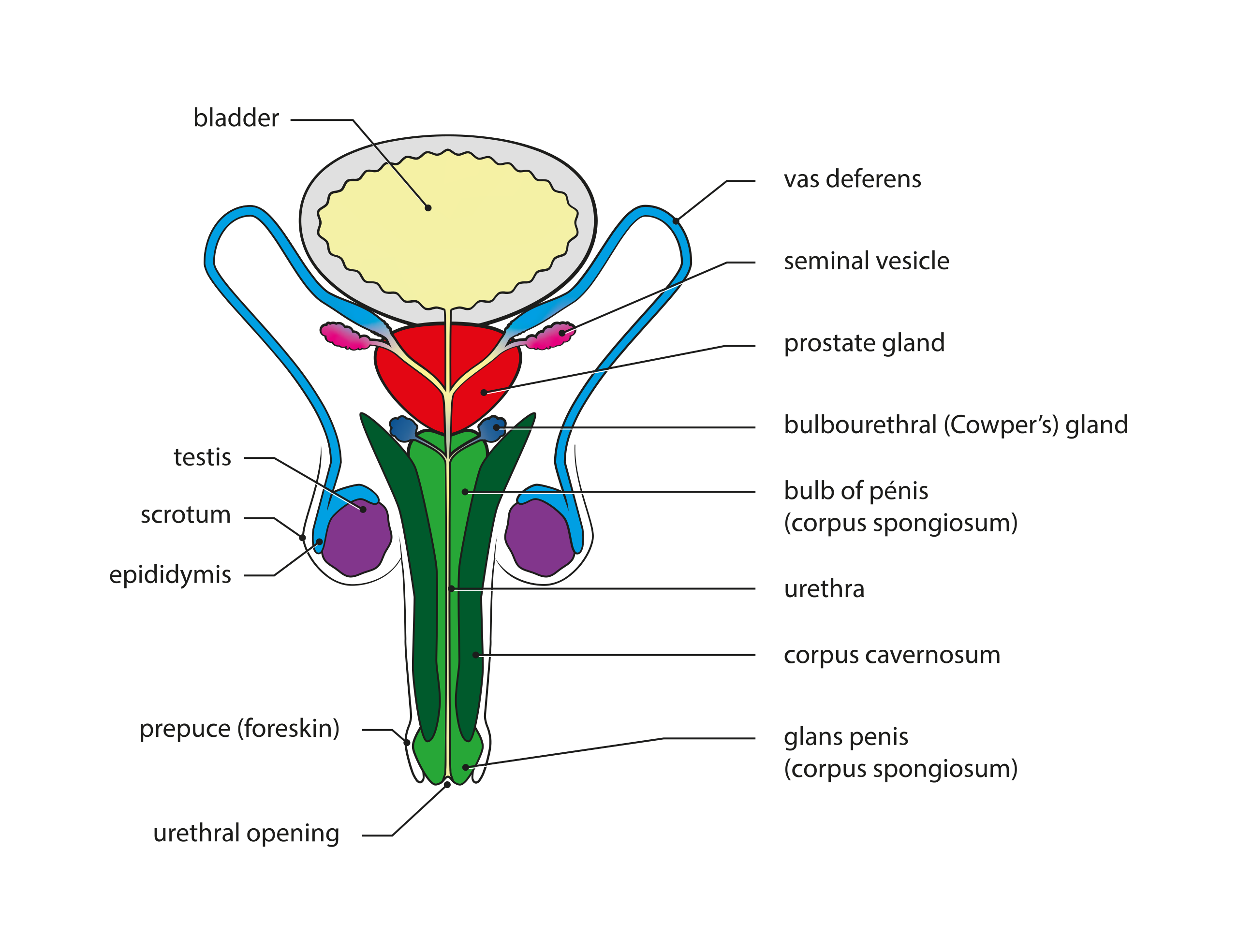
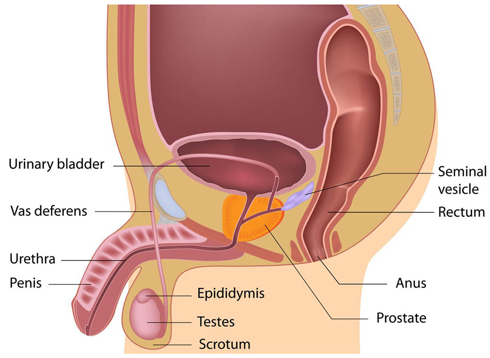
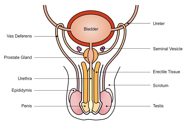
Components of Human Male Reproductive System
1.Testes
- Seminiferous tubules produce sperms .
- Interstitial cells produce testosterone
2. Ducts
- Epididymis
- Vas Deferens
- Ejaculatory Duct
3. Accessory Glands
Seminal Vesicle
Prostate Gland
Bulbo-Urethral Glands
4. Supportive Structure
Penis-
Transfers Sperms into the female Vagina
Testes
- Paired, oval-shaped male sex organs.
- Consist of seminiferous tubules, where the sperms are produced.
- Produce a male sex hormone called testosterone.
- Testosterone brings about changes in appearance of boys at puberty.
Also Check – Female Reproductive System
Table of Contents
Location of testes
The two testes (popularly called testicles) are oval organs which are contained in a thin-walled sac of skin called scrotum.
In the embryonic stage, the testes are contained within the abdomen.
They descend into the scrotum shortly before birth.
An abnormal condition results when they do not descend and it leads to sterility, i.e., incapability to produce sperms.
A slightly higher temperature inside the body does not permit maturation of sperms. But in the normal condition, being in a separate sac suspended from the body, the testes escape too much body heat.
Temperature regulation in the testes
Sperms are produced in the testes at a temperature 2 to 3°C lower than that of the body. This temperature is regulated in a strange manner through the movements of the scrotum wall.When it is too hot, the skin of the scrotum loosens so that the testes hang down away from the body. When it is cold, the skin contracts in a folded manner and draws the testes closer to the body for warmth.
Structure of testis
Each testis is encased in a capsule which is internally partitioned into around 250 lobules. Each lobule contains :
(i) Seminiferous tubules
- In Seminiferous tubules the sperms are produced.
- The process is called spermatogenesis.
(ii) Interstitial cells
- They are the packing tissues between the coils of the seminiferous tubules. The interstitial cells also called Leydig cells produce the male hormone testosterone.
- The sperm producing cells of the seminiferous tubules keep multiplying and produce sperms.
- The mature sperms pass into a small network of tubes.
- From the network, arise 12-14 ducts which join a small tubular knot, the epididymis fitting like a cap on the upper pole of the testis.
- The epididymis is continued by the side of the testis upto its back from where a distinct tube sperm duct (vas deferens) arises.
- The epididymis internally contains a single coiled tube which traverses from the upper part of the testis to its back and then it continues into the sperm duct.
- The epididymis stores the sperms for some days during which they mature and become motile (capable of moving).
Sperms
• Tiny and motile bodies that use their long tail to move through the female reproductive tract.
Scrotum
Small pouch that contains testis.
Present outside the abdominal cavity.
As sperms are formed here, this requires a lower temperature than the normal body temperature,
Vas deferens
Tube-like structure which connects testis to the urethra in order to allow the passage of semen.
It is also called as Sperm Ducts.
Vas deferens from each testis travels upward into the abdomen passing through an Inguinal canal.
The inguinal canal originally is the one which allows the descent of testes along with their ducts, blood vessels, nerves, etc.
Sometimes, due to pressure in the abdomen, the intestine bulges into the scrotum through the inguinal canal and causes the most common type of hernia.
The two sperm ducts loop over the ureters of their side, come together, and join the median duct, or urethra, at the back of the urinary bladder.
ACCESSORY GLANDS
Three male accessory glands are as follows
- A pair of lobulated glands located between the posterior surface of the urinary bladder and the rectum.
- A duct from each seminal vesicle joins the corresponding sperm duct just before it unites with the urethra.
- It produce a secretion which serves as a medium for the transportation of the sperms.
- The mixture of this fluid and the sperms produces la milky fluid, the semen. In the sperm duct, the sperms are sluggish, but by the addition of this secretion, they become active.
- A bilobed structure which surrounds the urethra close to its origin from the bladder.
- It pours an alkaline secretion into the semen as it passes through the urethra.
- It neutralizes acid in a female’s vagina.
- Bulbo-urethral glands (or Cowper’s glands)
- These are two small ovoid glands which open into the urethra just before it enters the penis.
- The secretion serves as a lubricant.
Also Check – Male Reproductive System Diagram
Urethra
- Common passage for both the sperms and urine.
- It never carries both of them at the same time.
PENIS
- The penis lies in front of the scrotum, cylindrical in shape, serves for the passing out of both semen and urine .
- It is a highly vascular organ, having erectile tissues and vascular spaces. Under the influence of sexual stimulation, blood flows in large amount into the penis and enters into the vascular spaces or sinuses, which makes it rigid and erect.
- Expansion of these spaces compresses the veins and thus blood cannot drain out.
- Such a condition of penis is called erection.
- Transfers sperms into the vagina of the female during copulation.
Semen
Semen is the mixture of sperms and secretions from seminal vesicles, prostate and Cowper’s glands.
It is a milky fluid. Its average amount is 2-3 ml in a single
ejaculation and contains 200,000,000-400,000,000 sperms.
4 Comments on “Male Reproductive System Class 10”