Table of Contents
Cell Division
Cell division is the process by which a cell divides into two or more daughter cells.
There are two main types of cell division:
- Mitosis
- Meiosis.
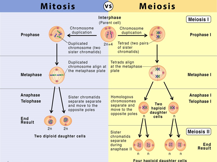
Mitosis is the process by which a single cell divides into two identical daughter cells. This is how cells replicate themselves during growth and tissue repair.
Meiosis is the process by which a single cell divides into four non-identical daughter cells. This is how cells produce gametes, or reproductive cells, such as sperm and eggs.
Both mitosis and meiosis involve a series of steps that involve the replication and separation of genetic material, as well as the division of the cytoplasm and cell membrane. Cell division is regulated by various proteins and enzymes that ensure that the process occurs accurately and efficiently.
Also Check – Mitosis In Plant Cell and Animal Cell- Differences and Similarities
Mitosis and Meiosis
Mitosis
- Mitosis is the process by which a cell divides its nucleus and cytoplasm into two identical daughter cells.
- During mitosis, the chromosomes in the nucleus are replicated and then separated into two sets, with one set going into each of the daughter cells.
- This ensures that each daughter cell gets a complete set of chromosomes and the same genetic information as the parent cell.
- The process of mitosis is essential for cell growth and repair, and it also plays a role in the development of multicellular organisms.
5 Stages of Mitosis Explained with Diagram
- Interphase
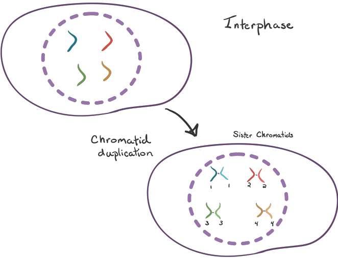
Interphase is the stage where the cell prepares for mitosis by replicating its DNA and synthesizing proteins.
- Prophase
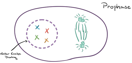
In the Prophase stage, the nucleolus disappears and the chromatin (uncoiled DNA) condenses into visible chromosomes. The mitotic spindle begins to form, and the centrosomes (organelles that organize the mitotic spindle) begin to move to opposite poles of the cell.
- Metaphase
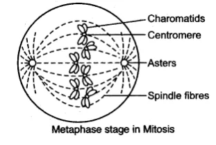
In Metaphase the chromosomes line up along the equator of the cell, with their centromere facing the center. The mitotic spindle is now fully formed and attached to the chromosomes at their centromere.
- Anaphase
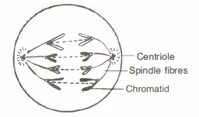
In Anaphase the centromere of each chromosome splits, and the two sister chromatids (half of the chromosome) are pulled towards opposite poles of the cell by the mitotic spindle.
- Telophase
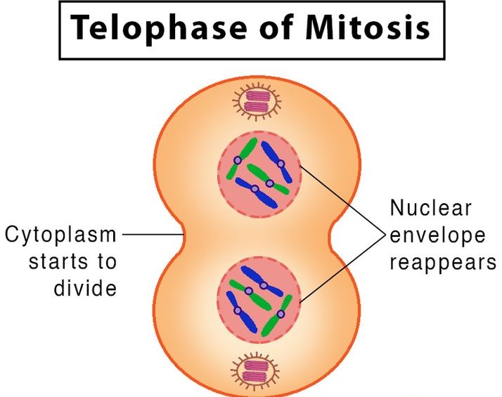
In Telophase the cell begins to divide into two daughter cells, with a new nuclear envelope forming around each set of chromosomes. A new nucleolus also appears.
- Cytokinesis
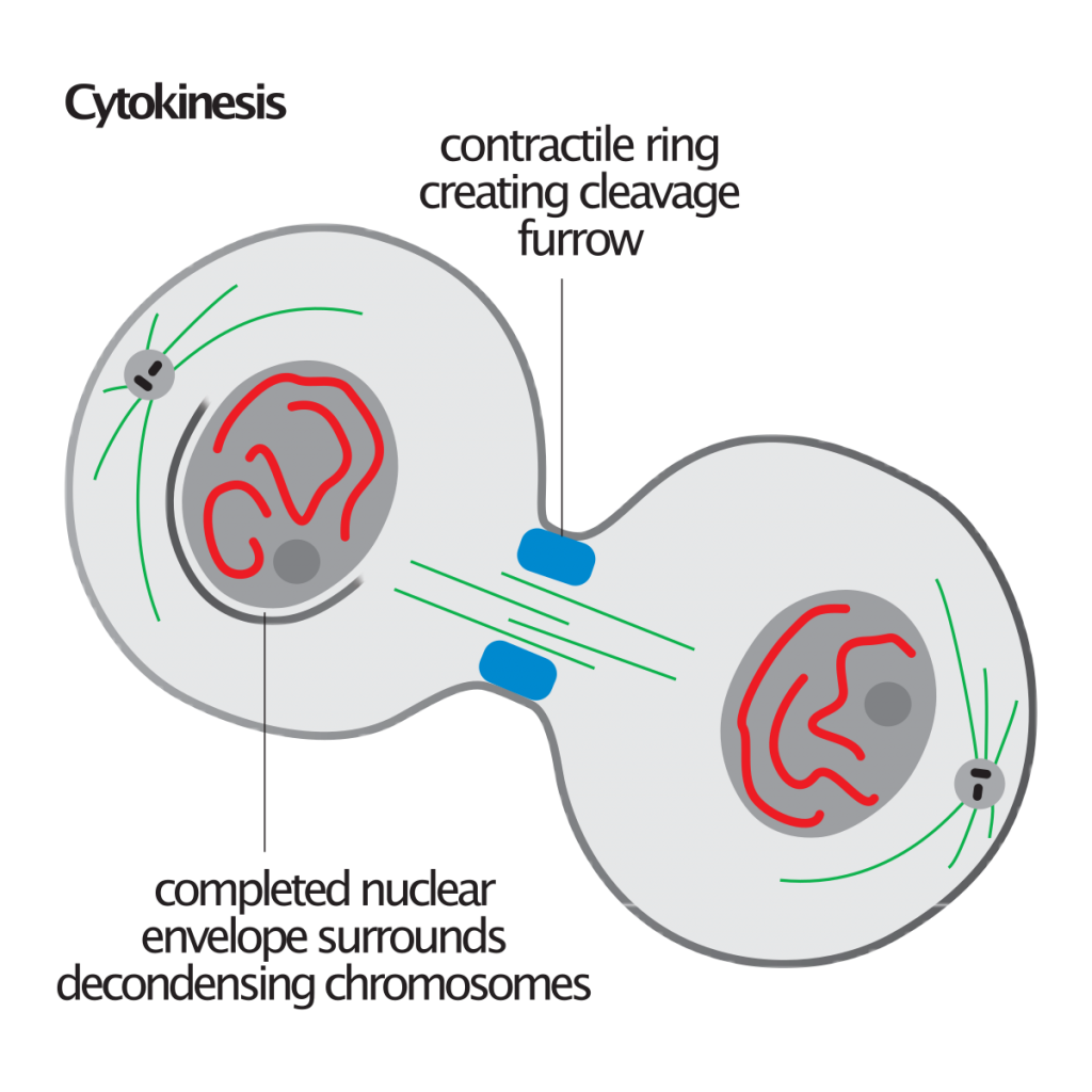
In Cytokinesis the cell’s cytoplasm divides, completing the process of cell division. Two new daughter cells are now formed.
Also Check – 7 Important Difference Between Mitosis and Meiosis
Also Check – 12 Important Significance of Mitosis
Also Check – Is Meiosis part of the Cell Cycle?
Meiosis
- Meiosis is a type of cell division that results in the production of four genetically unique daughter cells, each with half the number of chromosomes as the parent cell.
- Meiosis occurs in organisms that reproduce sexually and is essential for the proper segregation of genetic material during gamete (sperm or egg) production.
- During meiosis, the DNA of the parent cell is replicated, and then the chromosomes are separated into different daughter cells during two consecutive cell divisions.
- Meiosis results in the production of genetically unique cells that can combine with another gamete during fertilization to form a new individual.
- The daughter cells produced by Meiosis are Genetically Diverse because of the exchange of genetic material that occurs during the process of recombination. This diversity is important for the survival of species because it allows for the production of offspring with a wide range of traits, increasing the chances that some will be well-suited to survive in their environment.
Also Check – 5 Important Significance of Meiosis
Stages of Meiosis Explained with Diagram
There are two Stages of division during meiosis
- Meiosis I
- Meiosis II
During meiosis I, homologous chromosomes (pairs of chromosomes that contain the same genes) are separated, and during meiosis II, the sister chromatids (identical copies of a chromosome) of each chromosome are separated.
5 Stages of Meiosis 1 Explained with Diagram
- Interphase
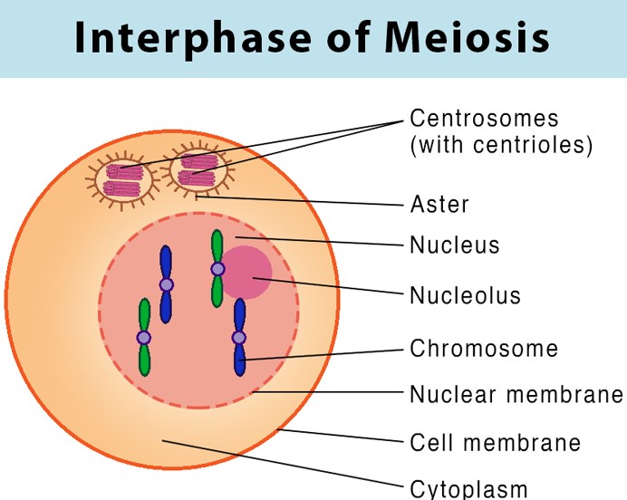
This is the preparatory stage for meiosis and is similar to interphase in mitosis. The cell grows, replicates its DNA, and prepares for cell division.
- Prophase I
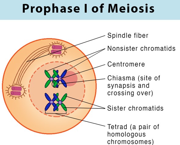
During this stage, the chromosomes become visible and start to condense. The nuclear envelope breaks down, and the centrosomes begin to move to opposite ends of the cell.
- Metaphase I
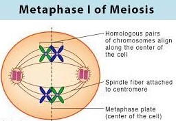
During metaphase I, the homologous chromosomes line up at the equator of the cell.
- Anaphase I
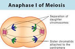
In anaphase I, the homologous chromosomes are separated and move to opposite poles of the cell.
- Telophase I
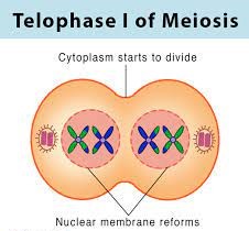
Telophase I is the final stage of meiosis I. The cell completes division, and two daughter cells are produced, each with half the number of chromosomes as the parent cell.
Also Check – What is the Importance of DNA copying in Reproduction
4 Stages of Meiosis 2 Explained with Diagram
- Prophase II
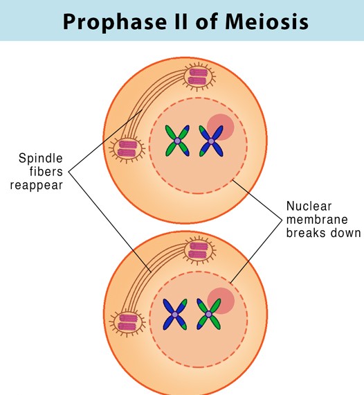
Prophase II is similar to prophase I, but the cells now have half the number of chromosomes as the parent cell.
- Metaphase II
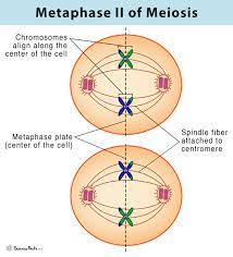
During metaphase II, the sister chromatids of each chromosome line up at the equator of the cell.
- Anaphase II
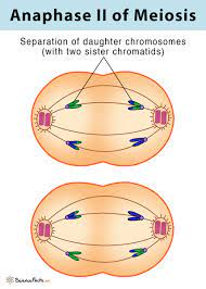
In anaphase II, the sister chromatids are separated and move to opposite poles of the cell.
- Telophase II
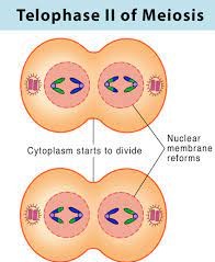
Telophase II is the final stage of meiosis II. The cell completes division, and four genetically unique daughter cells are produced, each with half the number of chromosomes as the parent cell.
Cytokinesis in Meiosis
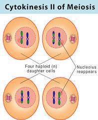
In meiosis, cytokinesis occurs after meiosis I and meiosis II, resulting in the production of four genetically diverse daughter cells.During both meiosis I and meiosis II, cytokinesis occurs, resulting in the production of four genetically diverse daughter cells.
Frequently asked Questions on Mitosis and Meiosis
What is the difference between Mitosis and Meiosis?
How does mitosis contribute to the growth and repair of tissues?
What are the five stages of mitosis and what happens during each stage?
Why is genetic diversity important for the survival of a species and how is it achieved during meiosis?
What are the two stages of division during meiosis and what happens during each stage?
How is meiosis different from mitosis in terms of the number of daughter cells produced?
What is the significance of DNA copying in reproduction?
What are homologous chromosomes and when are they separated during meiosis?
How does cytokinesis complete the process of Cell division?
How is the process of cell division regulated by proteins and enzymes?
7 Comments on “Mitosis and Meiosis”