What are Stomata?
Stomata are small openings, usually on the underside of a leaf, that allow the exchange of gases needed for photosynthesis. These openings are sensitive to light and close at night to conserve water.
Table of Contents
Stomata Diagram
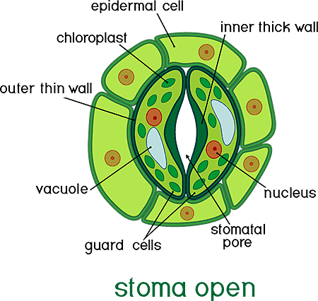
Stomata Diagram – Labels Explained
A typical stomata diagram contains the following labels:
Epidermal Cell – The outermost layer of cells on the surface of a leaf. It protects the underlying tissue from damage and helps regulate water loss.
Guard Cell – A pair of specialised cells that surround the stomatal pore and control its opening and closing. When the guard cells are turgid, the stomata pore opens and allows gas exchange and Transpiration. When the guard cells are flaccid, the pore closes and reduces water loss.
Stomatal Pore – The opening between the two guard cells that allows gas exchange and Transpiration. The size of the pore is regulated by the turgidity of the guard cells.
Also Check -8 Important Functions of Stomata
Inner Thick Wall – The inner wall of the guard cell, which is thicker than the outer wall. It provides structural support and helps maintain the shape of the guard cell.
Outer Thin Wall – The outer wall of the guard cell, which is thinner than the inner wall. It is more flexible and allows for changes in cell shape as guard cells open and close.
Chloroplast – An organelle within the guard cell that contains chlorophyll and is responsible for photosynthesis. Chloroplasts convert light energy into chemical energy used for cellular processes.
Vacuole –A large fluid-filled organelle in the guard cell that helps regulate the turgidity of the cell. When the vacuole is filled with water, the guard cell becomes turgid and the stomatal pore opens. When the vacuole is empty, the guard cell becomes flaccid and the pore closes.
Nucleus – The control centre of the cell that contains the genetic material and regulates cellular processes. The nucleus of the guard cell is responsible for regulating the expression of genes that control the opening and closing of the stomata.
Also Check- Mechanism of opening and Closing of the Stomata
The Stomata are surrounded by two curved guard cells that change shape to open and close the tiny pore. The inner thick wall of the guard cell provides structural support and helps maintain the shape of the cell, while the outer thin wall is more flexible and allows for changes in cell shape as the guard cells open and close. The chloroplasts in the guard cell are responsible for photosynthesis and the vacuole helps regulate the turgidity of the cell.
The lower surface of a leaf that is shaded from sunlight often has a higher number of stomata, so that less water is lost through transpiration. The role of stomata in transpiration is important because they allow the exchange of gases while regulating water loss.
Also Check – Role of Stomata in Transpiration
Diagram of Open and Closed stomata
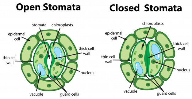
Stomata Closing
When conditions for photosynthesis are poor, such as when it is dark, water passes out of the guard cells. This makes them flaccid (limp) and causes the stomata to close. This also helps to stop water escaping when plant water levels are low.
Also Check- Protective Tissue in Plants-Define, Types, Functions
Stomata Opening
When plenty of light and water are available, water passes into the guard cells during the day by osmosis. This makes the guard cells turgid (swollen). They bend as they swell, causing the stoma to open.
8 Comments on “Diagram of Stomata”