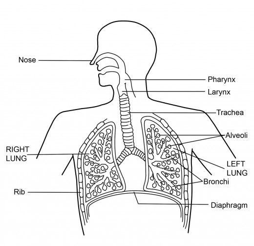
Human Respiratory System ( labeled in the diagram )
The parts of the Human Respiratory System ( labeled in the diagram )and their respective functions are as follow
Table of Contents
Nostrils
- Air is taken into the body through the nostrils.
- It is lined by fine hair and mucus which helps to filter the air entering through it.
Nasal Passage
- Air entering the nostrils leads to the nasal passage.
- It is mainly the conducting zone for air.
Pharynx
- Nasal chamber opens into the Pharynx.
- It passes air into larynx.
Larynx (or Sound Box)
- It is located in the neck region and in front of the trachea.
- It also produces sound.
Trachea (windpipe)
The air passes from the pharynx and goes into the trachea. Incomplete rings of cartilage keep the trachea open allowing the passage of air to the lungs. These ensure that theair passage does not collapse.
Also Check – Life Processes Class 10 MCQ
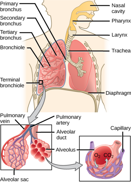
Bronchi
Trachea divides into two smaller tubes called bronchi after entering the thoracic cavity, which further extends into each lung.
Bronchioles
- Bronchi are subdivided into smaller tubes called bronchioles.
- Each bronchiole finally terminates into many alveoli.
Also Check – Mechanism of Respiration in Human Beings
Alveoli
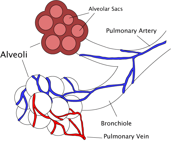
- These are balloon-like structures located inside the lungs.
- A large number of alveoli increases the surface area for the exchange of gasses.
- Human alveolar surface when spread can cover an area of 80 m².
Also Check – 12 Important Comparing factors between Alevoli And Nephrons with respect to their Location Structure and Functioning
Ribs
- These are 12 pairs of bones that form a cage in the thoracic region.
- Lungs and heart are safely placed in it.
- Movement of intercostal muscles attached to ribs helps in breathing.
Lungs
- These are primary organs for respiration, which arelocated on the two sides of the heart.
- These transport O₂ from the atmosphere into blood and release CO₂ from blood to the atmosphere.
- They are enclosed by the protective membranes called pleura.
Also Check – Importance Of Respiration
Diaphragm
- It is a muscular partition between the thorax and the abdomen and forms the base of the chest cavity.
- During inhalation, it flattens and increases the chest cavity.
Very nice 👍
Thank you So Much !