What is Mitosis?
The cells in animals and plants divide to produce new cells for growth or to replace cells that are damaged or worn out. This process of dividing to produce genetically identical cells is called Mitosis.
The process of cell growth and division is called the cell cycle.
Mitosis is the final stage of the cell cycle.
Mitosis produces two genetically identical cells and leaves the number of chromosomes unchanged.
Table of Contents
How Cells Divide in Mitosis
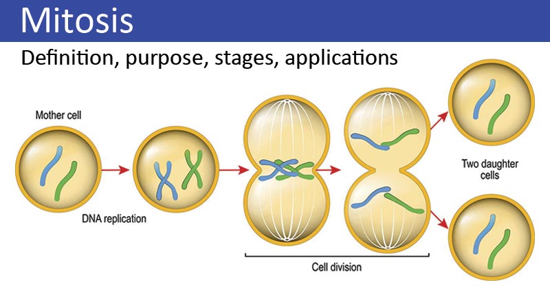
During mitosis, the nucleus of a cell divides to form two new nuclei, with two identical sets of chromosomes.
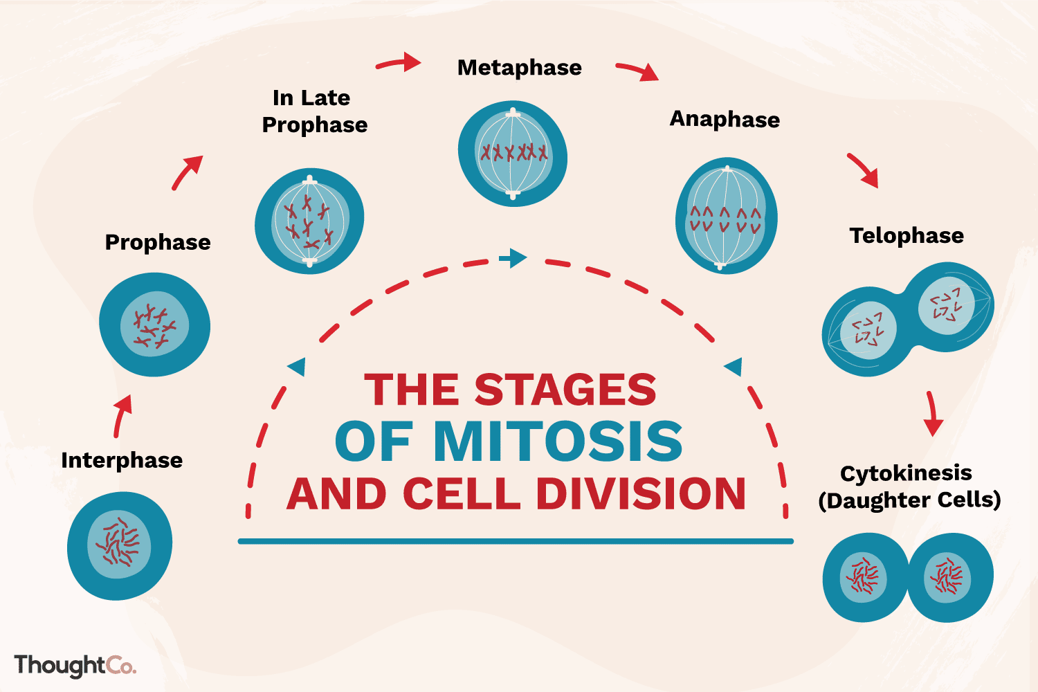
4 Stages of Mitosis
Mitosis has four stages
- Prophase
- Metaphase
- Anaphase
- Telophase
During the last stage of mitosis, the cytoplasm narrows between the two newly formed nuclei and the cell then splits to form two daughter cells.
Mitosis produces millions of new cells in the human body every second. Rapidly dividing cells are found in our skin, the roots of our hairs, and in the bone marrow, where new blood cells are made.
Mitosis Stages with Labeled Diagram
The DNA in a cell is stored in structures called chromosomes in the nucleus. Before mitosis, these look like long, tangled threads.
Prophase
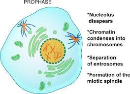
- The membrane enclosing the nucleus breaks up, and the DNA molecules wind up tightly, making the chromosomes condense (shorten and thicken).
- The chromosomes look X-shaped because each one is attached to an identical copy.
- Each side of the X is called a chromatid.
Metaphase
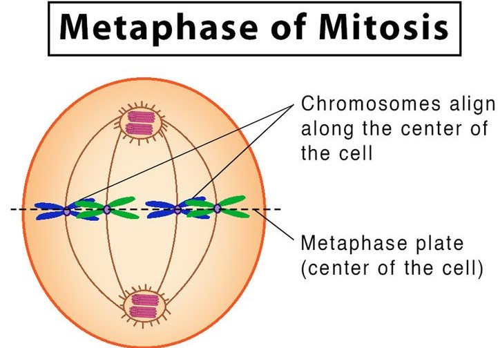
- The chromosomes are moved to the center of the cell by microscopic fibers (spindle fibers).
Anaphase
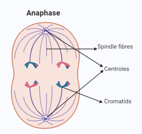
- The spindle fibers separate each chromosome into chromatids and pull set of chromatids to each end of the cell.
Telophase
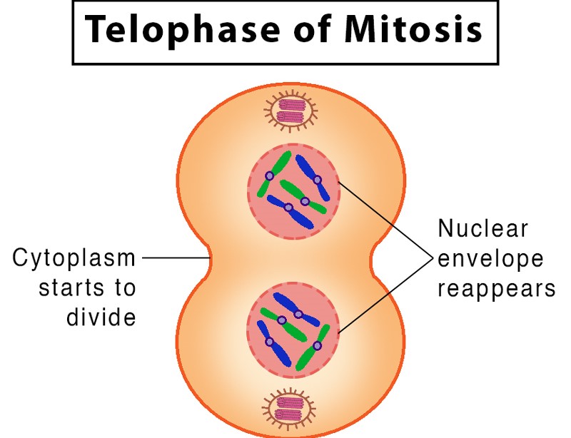
- Nuclear membranes form around each set of chromatids.
- The cytoplasm begins to divide (cytokinesis) to form two separate cells.
New cells
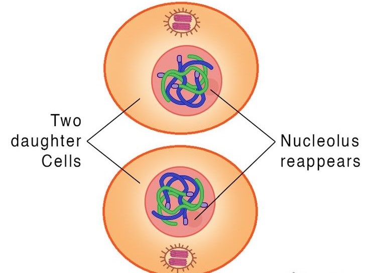
- Two identical daughter cells form and begin to grow.
- Before the cells are ready to divide again, the chromosomes will be copied to make duplicates of all the cell’s genes.
14 Comments on “Mitosis Explained with Diagram”