Salivary Glands Diagram

The salivary gland diagram provides a clear visual representation of the major salivary glands: parotid submandibular, and sublingual. Neatly labeled and easy to draw, this diagram helps you understand their anatomical location and functions. It is a valuable teaching tool for studying the important role of these glands in saliva production and oral health.

Salivary Glands Diagram Read More
Guard Cell Diagram

The diagram of the guard cells illustrates the specialised cells surrounding the stomata. These kidney-shaped cells with thicker outer walls swell when filled with water, causing the stomata to open. Conversely, they become flaccid when they lose water, causing the stomata to close. This process, controlled by vacuoles and cell walls, regulates gas exchange and water loss in plants.

Guard Cell Diagram Read More

Amoeba Diagram for Class 7

The Amoeba is a single-celled organism of Protozoa phylum.It is a type of unicellular Protist that can be found in a variety of environments. The amoeba diagram is a simple and easy-to-draw representation of this organism, neatly labeled to help students in class 7 understand its structure. The essential features of the amoeba diagram comprise the pseudopod, cytoplasm, food vacuole, nucleus and other parts.

Amoeba Diagram for Class 7 Read More

Human Circulatory System

The human circulatory system is responsible for delivering oxygen, nutrients, and hormones to cells and tissues throughout the body. It consists of the heart, blood vessels and blood. The heart pumps oxygenated blood to the body through arteries and the deoxygenated blood returns to the heart through veins. The circulatory system also helps remove waste products from cells and helps regulate body temperature.

Human Circulatory System Read More

Parts of Leaf – Structure, Types How Leaves Work , Function and Diagram

Leaves are an important part of plants. They come in different shapes and sizes, but all leaves have similar parts. The flat, green part of the leaf is called the blade and the stalk that attaches it to the stem is called the petiole. Leaves have tiny openings called stomata, which let in carbon dioxide and release oxygen. Leaves use sunlight to make food for the plant through a process called Photosynthesis.

Parts of Leaf – Structure, Types How Leaves Work , Function and Diagram Read More

Transverse Section of Leaf

The transverse section of a leaf is a cross-sectional view revealing the internal structure and organisation of cells, which is critical for understanding functions and adaptations.Leaf has several layers – the upper and lower epidermis, palisade and spongy parenchyma and vascular bundles. Stomata and air spaces regulate gas exchange, while xylem and phloem transport water, minerals, and nutrients.

Transverse Section of Leaf Read More

Xylem Diagram

A neatly labeled xylem diagram to understand xylem tissue. Xylem consists of vessel elements, tracheids, fibers and parenchyma cells each with unique functions in water and nutrient transport and structural support. Important features such as cytoplasm, the nucleus and bordered and simple pits are also shown in the diagram. Students can use this easy-to-draw diagram for their assignments and exams.

Xylem Diagram Read More

Phloem Diagram – Longitudinal and Transverse Section Diagram of Phloem Tissue

The Phloem is a plant tissue responsible for translocating organic nutrients and other substances from leaves to other parts of the plant. Longitudinal and Transverse Phloem Diagrams show the internal structure of the tissue, including Sieve tubes, Companion Cells, Phloem Fibers and Phloem Parenchyma cells.Netaly labelled easy to draw Phloem Diagram – Longitudinal and Transverse Section Diagram of Phloem Tissue explained with description of Labels

Phloem Diagram – Longitudinal and Transverse Section Diagram of Phloem Tissue Read More
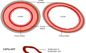
Neatly labelled easy to draw diagram of blood vessels - arteries ,veins, capillaries

Blood Vessels Diagram

The blood vessels diagram shows the structure of arteries and veins. Arteries have a thick tunica media layer to handle the high-pressure blood flow while veins have a thinner tunica media and a larger lumen to accommodate a higher volume of blood. the smallest blood vessels have thin endothelial cell walls and a narrow lumen that allows for the exchange of nutrients and waste products with pericytes and red blood cells. A neatly labeled diagram makes it easy to understand.

Blood Vessels Diagram Read More