Table of Contents
What is Simple Epithelial Tissue?
Definition-
Simple epithelial tissue consists of a single layer of cells that covers various body surfaces and lines internal cavities. Each cell is directly in contact with a supporting basement membrane, which regulates material exchange and provides structural support. This differs from stratified epithelial tissue, which has multiple layers of cells and is found in areas needing greater protection.
Basic Characteristics-
Simple epithelial tissue plays key roles in protection and substance exchange within the body. It forms the outer surface of the skin and lines internal cavities, including those of the respiratory, digestive, and reproductive systems, as well as blood and lymph vessels. The single layer of cells in simple epithelial tissue enables efficient transfer of materials, crucial for functions such as gas exchange in lung alveoli and filtration in kidney nephrons.
The thin, single-cell layer structure makes simple epithelial tissue delicate, suited to internal areas where less physical protection is needed but rapid absorption and filtration are essential. For instance, it aids in nutrient absorption in the digestive tract and facilitates oxygen-carbon dioxide exchange in the respiratory system. This illustrates the body’s adaptation to varying physiological demands through specialised epithelial cells.
Structure of Simple Epithelial Tissue
Simple epithelial tissue is composed of a continuous, tightly fitted layer of cells. The organisation and structural components of this tissue enable its crucial functions in absorption, secretion, and selective permeability.
Cell Arrangement and Membranes-
The cells in simple epithelial tissue are arranged in a single layer where each cell is directly attached to a basement membrane. This membrane is a thin, fibrous layer composed of proteins and polysaccharides secreted by the epithelial cells themselves. It provides structural support and anchors the epithelial tissue to the underlying connective tissue.
Surfaces and Junctions-
One surface of the simple epithelial cells is always exposed to either an internal or external environment, depending on the location within the body. This could be facing the inside of an organ, such as a stomach lining, or it could be exposed to outside elements, as in the skin. The opposite side of these cells is attached to the basement membrane.
The integrity and function of epithelial tissue are maintained through several types of specialised junctions-
- Tight junctions seal the spaces between cells, preventing leakage of materials and maintaining a barrier.
- Adherens junctions help to cement adjacent cells together, providing mechanical stability to the tissue.
- Gap junctions allow for the communication between cells through the exchange of ions and small molecules, facilitating coordinated functions.
Types of Epithelial Membranes-
Epithelial tissues form two main types of membranes-
- Mucous membranes (mucosa) contain goblet cells that produce mucus, aiding in lubrication and protection. These membranes line body cavities that open to the exterior, such as the respiratory and digestive tracts.
- Serous membranes line the closed internal cavities of the body, including the pleural and pericardial cavities. These membranes are primarily composed of simple squamous epithelium and secrete a lubricating serous fluid that reduces friction between internal organs.
Glandular Epithelium-
Glands in the body are predominantly made up of epithelial cells. These can be classified into-
- Exocrine glands, which include structures like sweat glands and goblet cells, secrete their products into ducts that open onto an epithelial surface.
- Endocrine glands are ductless and release their products, typically hormones, directly into the bloodstream or surrounding interstitial fluid.
This structural organisation allows simple epithelial tissue to perform effectively across various locations in the body, from providing a barrier to facilitating absorption and secretion processes.
Also Check-5 Key Difference between Simple Tissue and Complex Tissue
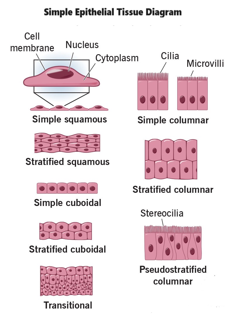
Simple Epithelial Tissue Diagram

Location of Simple Epithelial Tissue
Simple epithelial tissue is strategically located throughout the body to optimise its functions of absorption, secretion, and filtration. Here are the primary locations where simple epithelial tissue is found-
Body Cavities-
Simple epithelium lines several major body cavities, providing a smooth, frictionless interface. This includes-
- Peritoneal cavity- The lining of the abdominal cavity.
- Pleural cavity- The lining around the lungs.
- Pericardial cavity- The lining around the heart.
Blood and Lymph Vessels-
Simple epithelium forms the endothelium, the inner lining of all blood vessels and lymphatic vessels. This lining is crucial for managing the exchange of substances between the bloodstream or lymph and surrounding tissues.
Heart-
The inner lining of the heart, known as the endocardium, is composed of simple squamous epithelium. This smooth lining reduces friction, allowing for easier heart contractions and blood flow.
Respiratory System-
In the respiratory system, simple squamous epithelium lines the alveoli, the tiny air sacs in the lungs. This thin epithelial lining is essential for the efficient exchange of oxygen and carbon dioxide during the process of breathing.
Each location of simple epithelial tissue is specifically suited to the local environmental conditions and the physiological functions required in that area, emphasising the tissue’s role in protection, secretion, and absorption.
Functions of Simple Epithelial Tissue
Simple epithelial tissue plays several vital roles in the body, each related to its structure and location. Here are the key functions of simple epithelial tissue-
Protection-
- Simple epithelial tissue acts as a barrier against mechanical injuries, chemical exposure, fluid loss, and infections. For example, ciliated epithelium in the upper respiratory tract traps dust and pathogens, preventing them from entering deeper parts of the respiratory system.
Absorption-
- This tissue type is crucial in the absorption process. In the digestive tract, simple columnar epithelial cells absorb essential nutrients and water, facilitating their entry into the body’s circulation.
Filtration-
- Simple squamous epithelium, with its flat and thin cell structure, is ideally suited for filtration functions. This is observed in the kidneys where it forms part of the filtration barrier in nephrons, and in the lungs’ alveoli where gas exchange occurs.
Secretion-
- Epithelial cells are integral components of various glands. Simple cuboidal epithelium, found in glandular ducts, is involved in the secretion of enzymes, hormones, and other substances. Pseudostratified columnar epithelium, found in the respiratory tract, secretes mucus that traps foreign particles and bacteria, which are then expelled from the body.
Exchange of Substances-
- Epithelial tissue regulates the exchange of substances between the body and the external environment, as well as internally between different body parts. This includes controlling the entry and exit of substances across the epithelial barrier.
Sensation-
- Epithelial tissue also contains sensory receptors in specific areas such as the skin, nose, and taste buds. These receptors are essential for the sensation of touch, smell, and taste, providing critical information to the brain about the external environment.
Surface Specialisation-
- Adaptations like microvilli and cilia on epithelial cells enhance their functional capabilities. Microvilli increase the surface area for absorption, while cilia aid in moving mucus and other substances across the epithelial surface, particularly in the respiratory tract.
Types of Simple Epithelial Tissue
Simple epithelial tissue is categorised into several types based on the shape and arrangement of the cells. Each type is specialised for certain functions and locations within the body.
Simple Squamous Epithelium-
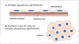
- Description- Composed of flat and thin cells, this type of epithelium facilitates the passive diffusion of gases and other substances.
- Function- Ideal for areas requiring rapid diffusion or filtration.
- Location- Found in the alveoli of the lungs, lining of the heart (endocardium), blood vessels (endothelium), and body cavities such as the peritoneal, pleural, and pericardial cavities.
Simple Cuboidal Epithelium-
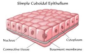
- Description- Consists of cube-shaped cells with roughly equal height and width, central spherical nuclei, and sometimes microvilli on the surface.
- Function- Primarily involved in secretion and absorption; forms the ducts and secretory portions of small glands.
- Location- Present in the kidney tubules, small ducts of glands, surface of the ovaries, and parts of the eye and thyroid.
Simple Columnar Epithelium-
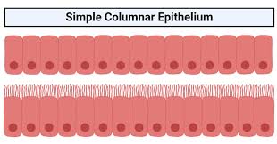
- Description- Characterised by tall, column-like cells with nuclei generally positioned near the base. These cells may have a striated border with microvilli to increase absorption surface.
- Function- Specialised for absorption and secretion; provides a protective barrier.
- Location- Lines most of the digestive tract (stomach to rectum), gallbladder, and excretory ducts of some glands. Ciliated varieties are found in the bronchi, uterine tubes, and uterus.
Pseudostratified Columnar Epithelium-
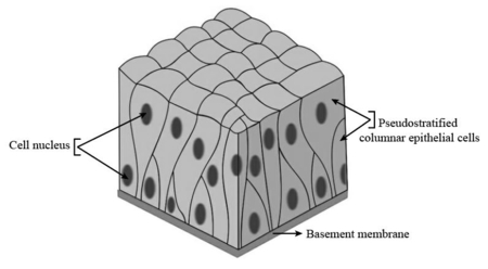
- Description- Appears layered due to nuclei at various depths but is actually a single layer of cells differing in height.
- Function- Mainly involved in secretion and movement of mucus by ciliary sweeping.
- Location- Lines the respiratory tract (nasal cavity to trachea) and portions of the male reproductive tract. The ciliated type helps in moving mucus out of the respiratory airways.
Table of Types of Simple Epithelial Tissue with location Structure and Function
| Type of Epithelial Tissue | Location | Structure | Function |
|---|---|---|---|
| Simple Squamous Epithelium | Blood vessels, heart (endocardium), alveoli, body cavities (peritoneal, pleural, pericardial) | A single layer of flat cells | Facilitates rapid diffusion or filtration; minimal protection required |
| Simple Cuboidal Epithelium | Kidney tubules, small ducts of glands, ovaries, parts of the eye and thyroid | A single layer of cube-shaped cells, sometimes with microvilli | Absorption and secretion; forms ducts and secretory parts of glands |
| Simple Columnar Epithelium | Digestive tract (stomach to rectum), gallbladder, excretory ducts of some glands, bronchi, uterine tubes, uterus | A single layer of tall, columnar cells, often with microvilli or cilia | Protection, absorption, secretion of mucus; ciliated types help move substances |
| Pseudostratified Columnar Epithelium | Respiratory tract (nasal cavity to trachea), parts of male reproductive tract | Appears layered due to nuclei at different heights but is a single layer of cells of varying height | Secretion, particularly of mucus; movement of mucus by ciliary action |
| Keratinized Epithelium | Skin (epidermal layer) | Multiple layers of epithelial cells, upper layers are dead and filled with keratin | Provides protection against abrasion, pathogens, and water loss; surface is waterproof |
| Transitional Epithelium (Urothelium) | Urinary bladder, ureters, part of the urethra | Stratified epithelium; cells change shape from cuboidal to flat when stretched | Allows the urinary organs to stretch and expand; acts as a barrier to prevent the reabsorption of toxic substances |