The Human Alimentary Canal, also known as the digestive tract or gastrointestinal tract, is an important structure in the body responsible for digesting and absorbing food. When we eat food, it is broken down into smaller components by different digestive enzymes and acids in different parts of the digestive tract. These smaller components are then absorbed into the bloodstream and transported to different parts of the body to provide energy and nutrients.
Table of Contents
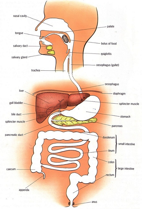
Human Alimentary Canal Diagram
Description of Human Alimentary Canal Diagram labels
Mouth- The mouth is the first part of the digestive system through which food enters the alimentary canal. It contains important organs such as the tongue, teeth and salivary glands.
Also Check – Explain the Role of Mouth in Digestion of Food
The Tongue- A highly muscular sensory organ located at the base of the buccal cavity. It carries several taste buds and helps mix food with saliva. In addition, it is also helpful in speech production.
Also Check – Digestive Glands – Definition , Types and Functions
Salivary Glands- These glands produce saliva, a digestive juice that contains enzymes such as amylase that help break down carbohydrates.
Also Check – 9 Important Functions of Salivary Glands
Teeth- Hard structures on the bones of the upper and lower jaws that are used to grind, cut and chew food.
Pharynx- A small funnel-shaped chamber behind the oral cavity that communicates with both the oesophagus and the trachea.
Oesophagus- A thin and long muscular tube leading into the stomach.
Stomach– The most dilated J-shaped part of the alimentary canal situated between the oesophagus and the small intestine below the diaphragm.It serves as a storehouse of food where partial digestion takes place through secretion of the gastric glands.The muscular walls of the stomach help to ensure that the food is well mixed with the digestive juices.The exit of food from the stomach is regulated by a sphincter muscle, which releases the food in small amounts into the small intestine.
Also Check – What is the role of Acid in our Stomach?
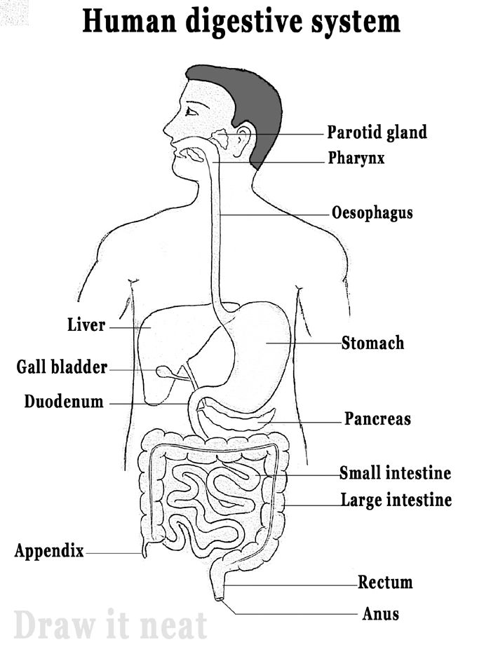
The Liver- The largest gland in the body that secretes bile juice.Bile emulsifies fats so that they can be digested more easily by enzymes.It is released into the small intestine through the bile duct.
Also Check – Process of Digestion in Human Beings
Gall Bladder- A small sac that stores the bile juices for further use.
Pancreas- A gland that secretes pancreatic juice containing enzymes such as amylase, trypsin and lipase.It is connected to the small intestine via its main duct, the pancreatic duct.
Also Check – 9 Important Function of Pancreas
Small Intestine– The longest part of the alimentary canal, fitted in a compact space due to extensive coiling.The small intestine is where the complete digestion of food into different components takes place.Secretions from the liver and pancreas enter the intestine to aid the digestive process.The inner lining of the small intestine has numerous finger-like projections called villi that increase the surface area for food absorption.
Also Check – Why do Herbivores have Longer Small Intestine than Carnivores
Large intestine- Although shorter, it is called a large intestine because it is larger in diameter than the small intestine.
Appendix– A small pouch-like structure attached to the large intestine.
Rectum- The last and wide chamber-like structure used to store faecal matter temporarily.
Anus- The end point of the digestive tract used to eliminate waste.
Also Check – How are Fats Digested in our Body ? Where does this process take place?
Also Check- How is the Small Intestine designed to absorb Digested Food ?
5 Comments on “Human Alimentary Canal Diagram Class 10”