Table of Contents
Placenta
Placenta is an organ that forms during pregnancy . It provides a vital link between the developing fetus and the mother’s blood supply. It is located in the uterus and is attached to the fetal side of the umbilical cord. Placenta acts as an interface between the mother’s and the fetus’s circulatory systems . It allows the exchange of oxygen, nutrients and waste products between the two. Placenta also produces hormones like human chorionic gonadotropin (hCG) that are important for maintaining pregnancy and supporting fetal development. After birth the placenta is expelled from the mother’s body .
Also Check – Structure Of The Placenta
Diagram of The Placenta
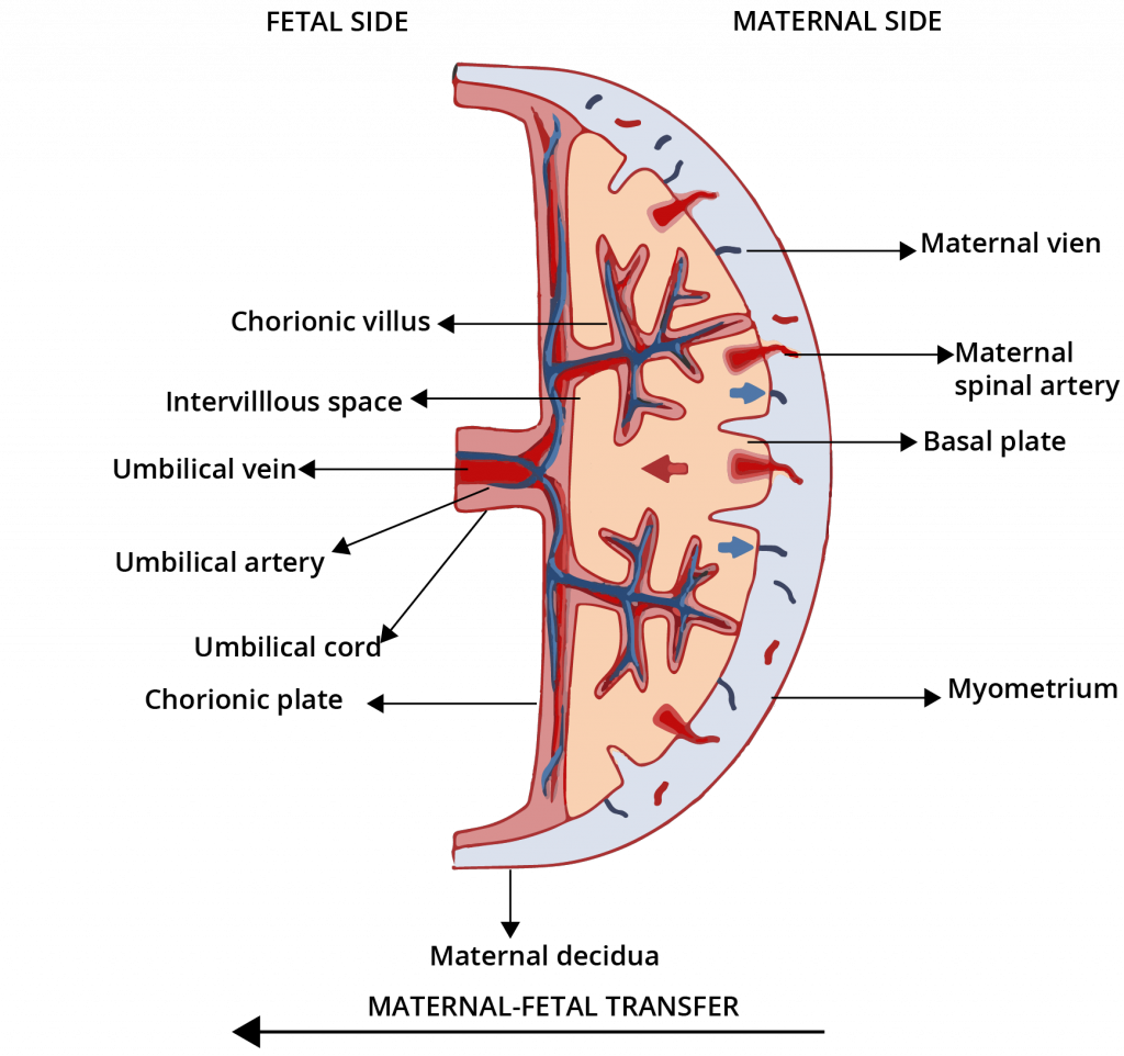
Description of Placenta Diagram labels
Description of Placenta Diagram labels is as follows –
Fetal side
Fetal side of the placenta is the surface that faces the fetus. It is covered by a layer of fetal membranes including the amniotic sac . Amniotic sac protects the fetus and provides a fluid-filled environment for it to grow. The fetal side of the placenta is also in direct contact with the umbilical cord which carries blood and nutrients to and from the fetus.
Also Check – 9 Important Functions of Placenta
Chorionic villus
Chorionic villi are small finger-like projections that extend from the chorion. It is the outermost layer of the placenta. Chorionic villus play an important role in exchanging gasses, nutrients and waste between the fetus and the mother’s blood supply. The large surface area provided by the chorionic villi helps to increase the efficiency of these exchanges.
Intervillous space
Intervillous space is the area between the chorionic villi in the placenta. It is filled with maternal blood which provides the fetus with oxygen and nutrients. The intervillous space is also involved in the exchange of waste products such as carbon dioxide between the fetus and the mother’s blood supply.
Umbilical vein
The umbilical vein is one of two blood vessels in the umbilical cord. It carries oxygen-rich blood from the placenta to the fetus. This blood is then used by the fetus to support its growth and development.
Umbilical artery
The umbilical artery is one of two blood vessels in the umbilical cord. It carries deoxygenated blood from the fetus to the placenta where it is oxygenated. The oxygenated blood is then returned to the fetus via the umbilical vein.
Umbilical cord
Umbilical cord is a flexible tubular structure that connects the fetus to the placenta. It contains two arteries and one vein. It carries blood between the fetus and the placenta. The umbilical cord is covered by a protective layer of membranes and is an essential structure for the transfer of nutrients, oxygen and waste products between the fetus and the mother’s blood supply.
Also Check – How does the Embryo get Nourishment Inside the Mother’s Body
Chorionic plate
Chorionic plate is the inner surface of the placenta that faces the fetus. Chorionic plate is in direct contact with the intervillous space and is therefore involved in the exchange of gasses, nutrients and waste products between the fetus and the mother’s blood supply. The chorionic plate is also covered by a layer of fetal membranes. It protects the fetus and provides a fluid-filled environment for it to grow.
Maternal side
Maternal side of the placenta is the surface that faces the mother. It is in direct contact with the uterine wall and is supplied with blood by the maternal spinal artery and other blood vessels. The maternal side of the placenta is also involved in the exchange of gasses, nutrients and waste products between the fetus and the mother’s blood supply.
Maternal decidua
The maternal decidua is the layer of the endometrium that is modified and thickened during pregnancy to form the placenta. It provides a supportive environment for the placenta to attach and function properly. The maternal decidua also secretes hormones like human chorionic gonadotropin (hCG)that play an important role in maintaining pregnancy.
Maternal vein
Maternal vein is a blood vessel that carries oxygen-rich blood from the placenta to the mother
Maternal spinal artery
Maternal spinal artery is a blood vessel that supplies blood to the placenta. It runs along the spinal column and is one of several arteries that provide blood to the placenta. The maternal spinal artery helps to ensure that the placenta has a constant supply of oxygen and nutrients to support fetal growth and development.
Basal plate
Basal plate is the lower surface of the placenta. It is in direct contact with the uterine wall. It is composed of the maternal decidua and is responsible for anchoring the placenta in place within the uterus. The basal plate also helps to prevent the placenta from separating from the uterus which can cause serious complications during pregnancy.
Myometrium
Myometrium is the smooth muscle layer of the uterus. It is responsible for contracting during labor and delivering the baby. During pregnancy the myometrium is stretched as the uterus expands to accommodate the growing fetus but it remains relatively relaxed. This helps to prevent preterm labor and ensure that the fetus has enough room to grow and develop.
Did you find this article helpful? We’d love to hear your thoughts and suggestions in the comments!
3 Comments on “Diagram of The Placenta”