Salivary Glands - Definition, Types, Location, Size, Ducts, Diagram, Characteristics, Secretion, Structure and Function

Salivary Glands – Definition, Types, Location, Size, Ducts, Diagram, Characteristics, Secretion, Structure and Function

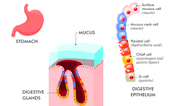
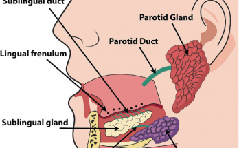
Salivary glands are exocrine glands responsible for saliva secretion. They have acinar cells for saliva synthesis and ductal cells for transport. Myoepithelial cells aid saliva movement. Major glands include parotid (largest), submandibular, and sublingual (smallest). Ducts are Stensen’s (parotid), Wharton’s (submandibular), and multiple (sublingual). Saliva lubricates, digests, and protects. Understanding their structure and function is vital.

Salivary Glands – Definition, Types, Location, Size, Ducts, Diagram, Characteristics, Secretion, Structure and Function Read More
Gastric Glands - Location , Structure, Types, Secretion and Functions

Gastric Glands – Location , Structure, Types, Secretion and Functions

Gastric glands, present in the stomach lining, play an important role in digestion. Structurally, they are made up of different cell types, including parietal, chief, and mucous cells. These glands secrete gastric juice, which consists of hydrochloric acid, pepsinogen and mucus. The secretions help break down food, sterilisation of ingested pathogens and absorb essential nutrients, thus fulfilling important functions in the digestive process

Gastric Glands – Location , Structure, Types, Secretion and Functions Read More

Salivary Glands Diagram

The salivary gland diagram provides a clear visual representation of the major salivary glands: parotid submandibular, and sublingual. Neatly labeled and easy to draw, this diagram helps you understand their anatomical location and functions. It is a valuable teaching tool for studying the important role of these glands in saliva production and oral health.

Salivary Glands Diagram Read More

Guard Cell Diagram

The diagram of the guard cells illustrates the specialised cells surrounding the stomata. These kidney-shaped cells with thicker outer walls swell when filled with water, causing the stomata to open. Conversely, they become flaccid when they lose water, causing the stomata to close. This process, controlled by vacuoles and cell walls, regulates gas exchange and water loss in plants.

Guard Cell Diagram Read More
peristalsis / perstaltic movement

Peristalsis-  The Rhythmic Journey Through Your Body

Peristalsis is a vital biological process that facilitates the movement of food through the digestive system. It involves rhythmic contractions of smooth muscles, creating a wave-like motion known as peristaltic waves. These waves propel food through the digestive tract and ensure efficient digestion and absorption of nutrients. Peristaltic movements are coordinated by the enteric nervous system and provide the motility necessary for proper gastrointestinal function.

Peristalsis-  The Rhythmic Journey Through Your Body Read More
emulsification of fats in digestion

Emulsification of Fats

Emulsification of fats is an important process in digestion in which large globules of fat are broken down into smaller droplets. This process is carried out by bile, a substance produced by the liver and stored in the gallbladder. Bile emulsifies fats by acting as a detergent, breaking down the fat into smaller droplets that can be easily digested and absorbed by the body. Without this process, the digestion and absorption of fats would be much less efficient.

Emulsification of Fats Read More

Transverse Section of Leaf

The transverse section of a leaf is a cross-sectional view revealing the internal structure and organisation of cells, which is critical for understanding functions and adaptations.Leaf has several layers – the upper and lower epidermis, palisade and spongy parenchyma and vascular bundles. Stomata and air spaces regulate gas exchange, while xylem and phloem transport water, minerals, and nutrients.

Transverse Section of Leaf Read More
How Amoeba Acquires its Food through the Process of Phagocytosis

How Amoeba Acquires its Food through the Process of Phagocytosis

Amoeba, a single-celled organism, acquires its food through the process of phagocytosis. During this process Amoeba uses pseudopodia to engulf its prey and forms a membrane-bound phagosome which fuses with lysosomes to form a phagolysosome. The prey is then digested. Factors such as temperature, pH and food availability affect the efficiency of phagocytosis in amoeba.

How Amoeba Acquires its Food through the Process of Phagocytosis Read More