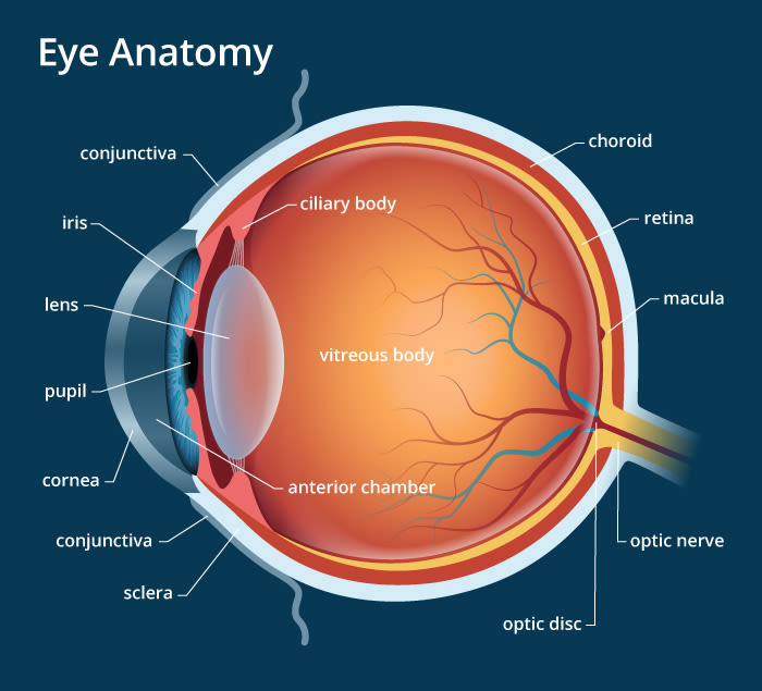
The main parts of the human eye are :
-
Cornea
-
Iris
-
Pupil
-
Ciliary muscles
-
Eye lens
-
Retina
-
Optic nerve
Table of Contents
Cornea :
- It is the transparent bulged out spherical membrane in the front of the eye.
- Light enters the eye through this membrane.
- Most of the refraction of light rays entering the eye occur at the outer surface of the cornea.
Lens :
- It is the central part of the eye that facilitates the image formation.
- It is a transparent, elastic, biconvex structure which is suspended in the cavity of the eyeball behind the pupil by means of suspensory ligaments.
- Lens is made of layers of non nucleated elongated cells and intracellular proteins.
- It is covered by a thin transparent membrane called lens capsule .
- The convexity of the lens is slightly more on the back as compared to the front.
- Stretching and relaxation of suspensory ligaments changes the focal length of the lens for accomodation .
Iris :
- It is an opaque muscular pigmented and perforated diaphragm having radial (dilator) and circular (sphincter) smooth muscles which are operated by sympathetic and parasympathetic nerves respectively.
- The iris provides colour to the eyes. It can be blue (only at the back), grey, brown (dark, light) or black (depending upon layers having pigment).
- The central perforation of the iris is called pupil.
- Its size is controlled by radial (contraction dilates pupil) and circular (contraction constricts pupils) muscle response to dim and strong light respectively.
Pupil :
It is a small hole between the iris through which light enters.
It opens up completely in the dim light due to contraction of iris muscles, but in bright light it becomes very small due to relaxation of iris muscles.
Ciliary Muscles :
They hold the lens in position and help in modifying the curvature of the lens.
Retina:
- It is the delicate inner nonvascular light sensitive coat of the eyeball.
- It is sensory and differentiated into two parts, outer pigmented part and inner nervous part.
- The pigmented part is made of cuboidal cells with dark brown granules and fringe-like protoplasmic processes. It continues beyond ora serrata.
Optic Nerve :
It transmits visual information from the retina to the brain.
Sclera :
- It is an opaque, fibrous, protective, outer layer of an eye containing and elastic fibre.
- It is also known as white of the eye.
Blind Spot:
It is the point at which the optic nerves leave the eye. It contains no rods and cones, so an image formed at this point is not sent to the brain.
Aqueous Humour :
Behind the cornea, we have a space filled with a transparent liquid called the aqueous humour
Vitreous Humour :
The space between eye lens and retina is filled with another liquid called vitreous humour.