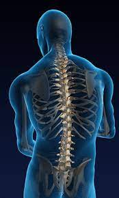
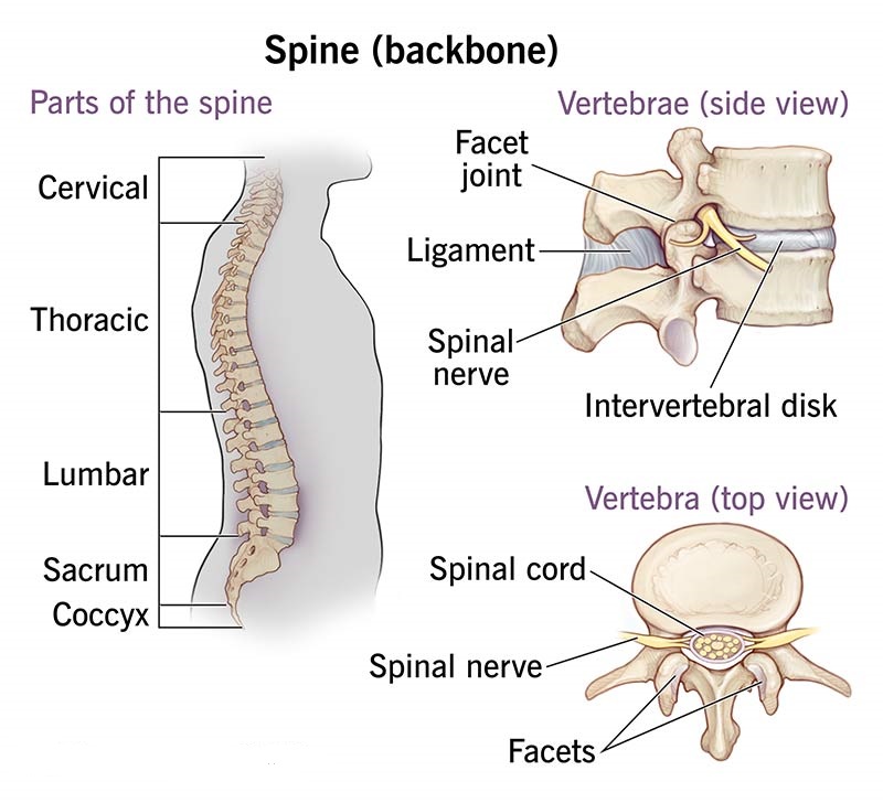
The spine, also known as the vertebral column, and the spinal cord are essential components of the human body’s anatomy. The spine is a series of approximately 33 bones called vertebrae, separated by intervertebral discs, which provide flexibility and cushioning. These bones are organised into five distinct regions- cervical, thoracic, lumbar, sacral, and coccygeal. Each region has unique structural features that support different functions such as movement, weight-bearing, and protection.
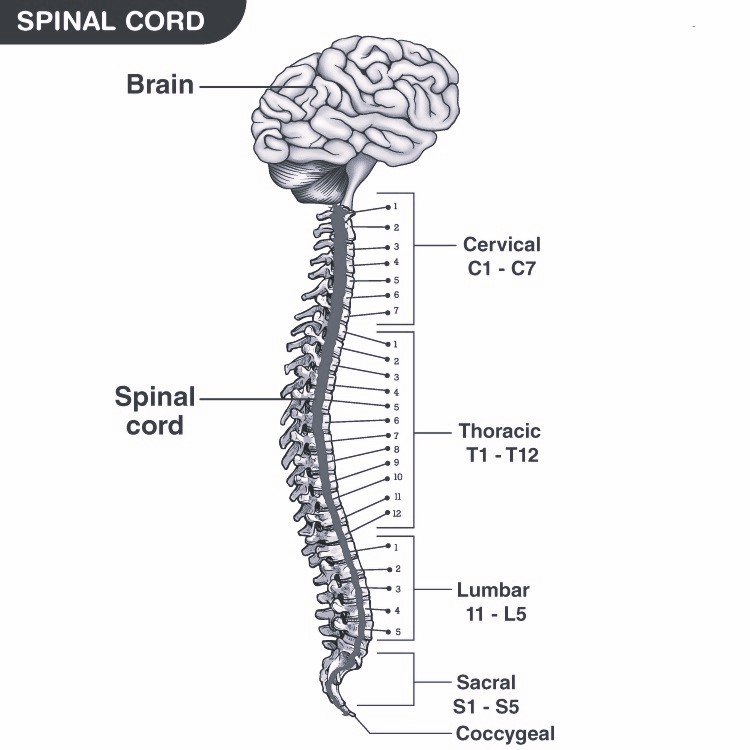
The spinal cord, part of the central nervous system (CNS), runs within the vertebral canal of the vertebral column. It extends from the foramen magnum at the base of the skull to the L1/L2 vertebra, where it terminates as the conus medullaris. The spinal cord is crucial for transmitting signals between the brain and the rest of the body and plays a key role in reflex actions.
Table of Contents
Anatomy of the Spine (Vertebral Column)
The vertebral column, commonly known as the spine, is structured into distinct regions, each with specialised vertebrae to support various functions and mechanical loads. Here we explore these regions in detail-
Cervical Vertebrae (C1-C7)
- Description and Function- The cervical vertebrae consist of the first seven vertebrae starting from the base of the skull. These are the smallest and lightest vertebrae, pivotal in supporting the head’s weight and facilitating its range of motion.
- Key Features for Motion and Support-
- Enhanced mobility for nodding and rotation movements.
- Unique vertebrae like the atlas (C1) and axis (C2) enable the head’s pivotal movements.
Thoracic Vertebrae (T1-T12)
- Description– Located in the chest region, these twelve vertebrae are characterised by their medium size and strength, with lateral facets for rib attachment, supporting the thoracic cage.
- Role in Upper Body Support-
- Structural support for the upper body and base for the rib cage.
- Limited mobility due to rib attachments, enhancing protective functions for vital organs.
Lumbar Vertebrae (L1-L5)
- Description- These five vertebrae are found in the lower back, known for their larger size and robustness, crucial for bearing the body’s weight.
- Key Characteristics-
- Thick and broad spinous processes for substantial muscle and ligament attachment.
- High resistance against bending and twisting forces.
Sacral Vertebrae (S1-S5)
- Description- In adults, these five vertebrae are fused to form the sacrum, a triangular bone that serves as a keystone for the pelvis.
- Connection to Pelvis-
- Integrates with the hip bones to form a stable pelvis-spine connection.
- Acts as a supportive structure for the upper body and a transmission point for lower body forces.
Coccygeal Vertebrae (Co1-Co4)
- Description- Comprising three to four fused vertebrae, they form the coccyx or tailbone, which is the terminal part of the vertebral column.
- Functional Relevance-
- Provides attachment points for ligaments and muscles of the pelvic floor.
- Acts as a support structure during sitting and provides leverage for muscles of the pelvic region.
Structure of a Vertebra
Each vertebra in the spinal column is meticulously structured to support and protect the spinal cord, while also allowing for movement and flexibility. Here’s a detailed look at the components-
Vertebral Body and Vertebral Arch
- Description- The vertebral body is the anterior, larger part of a vertebra and serves as the main weight-bearing structure. The vertebral arch is the posterior part that forms a protective canal for the spinal cord.
Role in Spinal Protection-
- Together, the vertebral body and arch enclose the vertebral foramen, providing a protective pathway for the spinal cord.
Bony Prominences
- Spinous Processes- Project posteriorly from the vertebrae, serving as attachment sites for muscles and ligaments.
- Transverse Processes- Extend laterally from each side of the vertebral arch, also providing muscle and ligament attachment points.
- Pedicles and Lamina- Bridge elements between the transverse and spinous processes, contributing to the architecture of the vertebral arch.
- Articular Processes- Protrude from the junctions of the pedicles and laminae, facilitating vertebral articulation with adjacent vertebrae.
Functions of the Vertebral Column
The vertebral column is crucial not just for structural support but also plays a key role in the overall functionality of the body-
- Protection- Encloses and safeguards the spinal cord within the spinal canal.
- Support- Bears the weight of the body above the pelvis, providing a central structure for body support.
- Axis Formation- Acts as the physical axis of the body, crucial for upright posture.
- Movement- Facilitates movement and flexibility in conjunction with muscles and ligaments.
Also Check – 6 Key Difference between Spine (Backbone) and Spinal cord
Anatomy of the Spinal Cord
General Structure
Position and Extent-
- The spinal cord is part of the central nervous system (CNS), situated within the vertebral canal of the vertebral column. It extends from the foramen magnum at the base of the skull to the L1/L2 vertebra, ending in the conus medullaris.
Division-
- Cervical Region- Neck area.
- Thoracic Region- Chest area.
- Lumbar Region- Lower back.
Components of the Spinal Cord
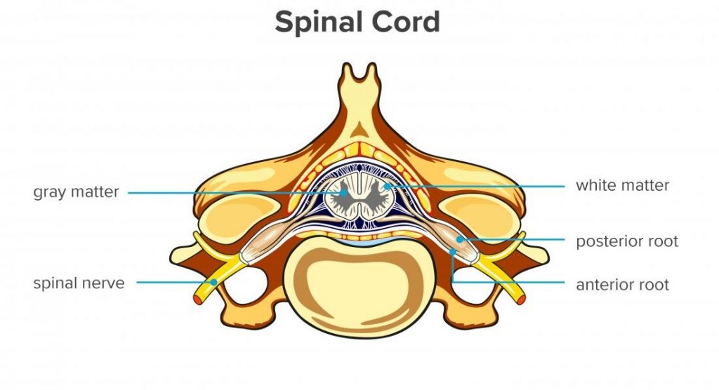
White Matter-
- The outer layer of the spinal cord, consisting of myelinated nerve fibres that carry signals to and from the brain.
Gray Matter-
- The central part of the spinal cord, shaped like a butterfly, containing neuronal cell bodies.
Horns-
- Anterior Horns- Contain motor neurons that send signals from the brain to the muscles.
- Posterior Horns- Contain sensory neurons that carry sensory information from the body to the brain.
Spinal Cord Functionality
- Conducting Impulses- The spinal cord transmits signals between the brain and the rest of the body.
- Reflex Actions- It generates reflexes, enabling quick, involuntary responses to stimuli.
Protective Coverings
- Meninges- Three layers of tissue that protect the spinal cord.
- Dura Mater- The tough, outermost layer.
- Arachnoid Mater- The middle layer, resembling a spider web.
- Pia Mater- The delicate, innermost layer that clings to the surface of the spinal cord.
Spinal Nerves
Structure and Function
Formation from Dorsal and Ventral Roots-
Each spinal nerve is formed by the merging of two types of roots-
- Dorsal Roots- Carry afferent (sensory) axons that transmit sensory information from the body to the spinal cord.
- Ventral Roots- Carry efferent (motor) axons that send motor signals from the spinal cord to the muscles.
Role in Sensory and Motor Signal Transmission-
- Sensory Function- Dorsal roots receive sensory information from various parts of the body and send it to the brain via the spinal cord.
- Motor Function- Ventral roots transmit motor commands from the brain to the muscles, enabling movement.
Distribution and Location
Number and Arrangement- There are 31 pairs of spinal nerves, each corresponding to specific vertebral levels-
- Cervical Nerves- 8 pairs (C1-C8)
- Thoracic Nerves- 12 pairs (T1-T12)
- Lumbar Nerves- 5 pairs (L1-L5)
- Sacral Nerves- 5 pairs (S1-S5)
- Coccygeal Nerves- 1 pair (Co1)
Location-
- Each spinal nerve exits the vertebral column through an opening called the intervertebral foramen, located between adjacent vertebrae.