The structural and functional differences between chromosome and chromatin is essential for students and researchers in genetics and molecular biology. Chromatin is the material that makes up chromosomes, consisting of DNA and proteins, and is involved in the compact packaging of DNA within the nucleus of cells. Chromosomes, formed from condensed chromatin, become visible and functionally significant during cell division, ensuring accurate distribution of genetic material. This article examines these components in detail, clarifying their roles and differences to enhance comprehension of cellular processes.
Table of Contents
Chromatin
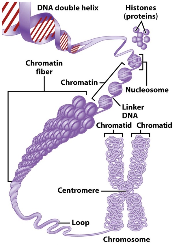
- Definition- Chromatin is the combination of DNA and proteins that forms the contents of the nucleus of a cell. It serves as the material out of which chromosomes are organised.
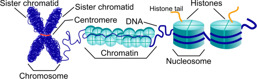
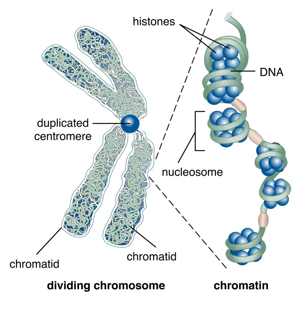
- Structure- Consists of nucleosomes, which include DNA wrapped around histone proteins, forming a fibre approximately 10 nm in diameter.
- Appearance- Appears as long, thin fibres that are loosely packed within the nucleus, visible under an electron microscope.
- Function- Chromatin facilitates DNA replication, RNA transcription, and DNA repair by allowing access to enzymes and other molecular machinery.
- Presence- Exists throughout the cell cycle, altering its condensation levels as needed for genetic activity.
Chromosome
- Definition- Chromosomes are distinct structures composed of condensed chromatin, visible during cell division, that carry genetic information in the form of genes.
- Structure- Higher order of DNA organisation, condensing DNA significantly to facilitate organised distribution during cell division.
- Appearance– Compact, thick, and ribbon-like during cell division, clearly visible under a light microscope.
- Function- Ensures accurate replication and distribution of genetic material during cell division, maintaining genetic stability.
- Presence- Prominently visible and highly condensed only during specific stages of cell division (metaphase, anaphase).
5 Key Differences between Chromosome and Chromatin in Tabular Format
| Aspect | Chromatin | Chromosome |
|---|---|---|
| Definition | DNA-protein complex within the nucleus. | Condensed chromatin visible during cell division. |
| Structure | Loose fibres of DNA wrapped around histones. | Highly condensed and organised DNA. |
| Appearance | Thin and fibrous, visible under electron microscopy. | Thick, compact structures visible under light microscopy. |
| Function | Supports DNA transcription, replication, and repair. | Ensures accurate DNA segregation during cell division. |
| Presence | Throughout the cell cycle, varying condensation. | Distinctly visible only during cell division stages. |
Differences between Chromosome and Chromatin – Detailed Explanation
Structure and Functionality-
- Chromatin- The structure of chromatin allows it flexibility to open or condense as needed, making it accessible for transcription and replication processes. This dynamic structure supports the cell’s metabolic and regulatory needs by modulating the exposure of DNA sequences.
- Chromosome- In contrast, the primary function of the chromosome structure is to be sufficiently compact during cell division to prevent tangling and ensure chromosomes are distributed evenly to daughter cells. This high level of organisation is crucial for the maintenance of genomic integrity.
Visibility and Identification-
- Chromatin- Due to its less condensed state, chromatin is not usually visible under light microscopy but can be seen with an electron microscope where its fibrous nature is apparent.
- Chromosome- Chromosomes, however, are easily identified under a light microscope during cell division due to their condensed state, which makes them appear as distinct entities.
Role in Genetics and Cellular Biology-
- Chromatin- By modulating its structure, chromatin plays a direct role in regulating gene expression. Its accessibility to transcription factors and RNA polymerases is crucial for the normal functioning of genes.
- Chromosome- Chromosomes are critical during the process of cell division, particularly during mitosis and meiosis, where they ensure that DNA is correctly replicated and each new cell inherits an accurate copy of the genome.
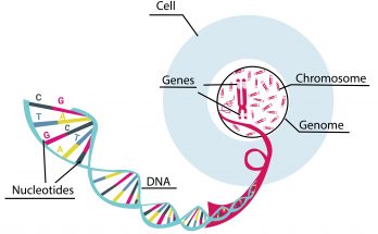
Also Check – 4 Important Differences Between Genes and Chromosomes
Also Check – Insights into Chromatin Structure and Dynamics in Plants
FAQs on Chromosome and Chromatin
What is chromatin and what is its primary function?
- Answer- Chromatin is a complex of DNA and proteins (mainly histones) found within the nucleus of a cell. Its primary function is to package DNA into a smaller volume to fit in the cell, control gene expression, and facilitate DNA replication and repair.
Describe the structure of a chromosome.
- Answer- A chromosome is a highly organised structure consisting of condensed chromatin. It is composed of DNA tightly coiled many times around proteins called histones. During cell division, chromosomes become visible under a light microscope as distinct entities.
Also Check – 6 Important Differences Between DNA and Chromosomes
How does the appearance of chromatin differ from that of chromosomes?
- Answer- Chromatin appears as thin, long fibres that are loosely packed within the nucleus and can only be seen clearly under an electron microscope. Chromosomes, however, are compact, thick, and ribbon-like structures that are easily observed under a light microscope during cell division.
What are the functional differences between chromatin and chromosomes?
- Answer- Chromatin supports the cell’s everyday metabolic functions, including transcription, replication, and DNA repair. Chromosomes, however, are primarily involved in ensuring accurate DNA segregation during cell division, maintaining genetic stability.
Also Check – 6 Key Differences Between DNA and Genes
When are chromosomes distinctly visible within the cell cycle?
- Answer- Chromosomes are distinctly visible during the metaphase and anaphase stages of cell division when they are most condensed.
Explain the importance of the structural organisation of chromosomes during mitosis.
- Answer- The structural organisation of chromosomes during mitosis is crucial for the even distribution of genetic material to daughter cells. This organisation helps prevent DNA tangling and breakage, ensuring that each daughter cell receives an accurate copy of the genome.
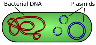
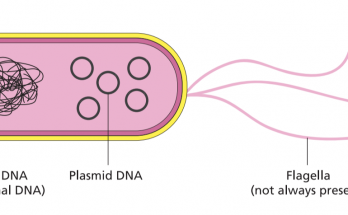
Also Check – 8 Key Differences Between Plasmid DNA and Chromosomal DNA
What role does the condensation of chromatin into chromosomes play in heredity?
- Answer- The condensation of chromatin into chromosomes during cell division ensures that genetic information is correctly replicated and evenly distributed among offspring, thus playing a key role in heredity.
Differentiate between euchromatin and heterochromatin.
- Answer- Euchromatin is a lightly packed form of chromatin that is accessible to transcription enzymes and is active in transcription, making it crucial for gene expression. Heterochromatin, on the other hand, is tightly packed, less accessible, and generally transcriptionally inactive, contributing to the structural integrity of the genome.
How can chromatin structure affect gene expression?
- Answer- The structure of chromatin affects gene expression by controlling the accessibility of DNA to transcription factors and RNA polymerase. When chromatin is loosely packed (euchromatin), genes are more likely to be expressed. Conversely, tightly packed chromatin (heterochromatin) can inhibit gene expression by restricting access to DNA.
What methods are used to visualise chromatin and chromosomes, and why are different methods used?
- Answer- Chromatin is best visualised using an electron microscope due to its less condensed state, while chromosomes, which are highly condensed, can be observed under a light microscope during cell division. These methods reflect the different levels of DNA compaction and are chosen based on the specific cellular structures being studied.